Page Contents
- 1 OVERVIEW
- 2 ORIENTATIONS USED FOR CHEST X-RAYS
- 3 QUALITY MEASURES FOR A CHEST X-RAY
- 4 ANATOMY ON CHEST X-RAYS
- 5 SYSTEMATIC APPROACH TO INTERPRETING A FRONTAL CHEST X-RAY: ABCDE APROACH
- 6 FRONTAL VIEW: AIRWAY
- 7 FRONTAL VIEW: BONES
- 8 FRONTAL VIEW: CARDIAC
- 9 FRONTAL VIEW: DIAPHRAM
- 10 FRONTAL VIEW: EVERYTHING ELSE
- 11 SYSTEMATIC APPROACH TO INTERPRETING A LATERAL CHEST X-RAY
- 12 LATERAL VIEW: RETROSTERNAL CLEAR SPACE
- 13 LATERAL VIEW: HILAR REGION
- 14 LATERAL VIEW: FISSURES
- 15 LATERAL VIEW: THORACIC SPINE
- 16 LATERAL VIEW: DIAPHRAM AND POSTERIOR COSTOPHRENIC SULCI
OVERVIEW
Interpreting a chest X-ray is a very valuable skill given how commonly it is ordered. The following guide helps walk you through a comprehensive approach to understand and interpreting chest X-rays. As a refresher read this guide on the fundamentals of X-ray studies to help learn some of the basics.
ORIENTATIONS USED FOR CHEST X-RAYS
Before delving too deep into the interpretation of chest X-rays, we must first learn about the different orientations in which the image can be taken. The orientations used for a chest X-ray include:
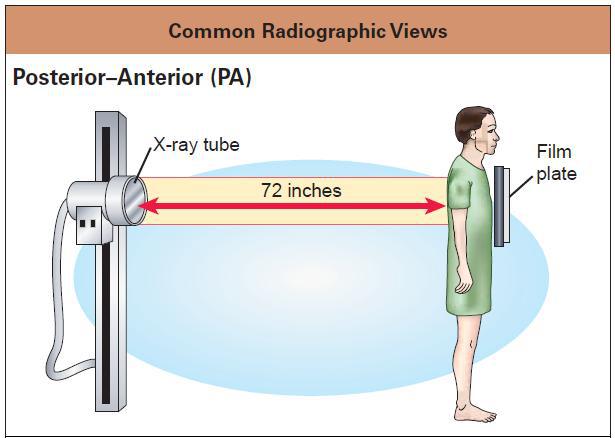
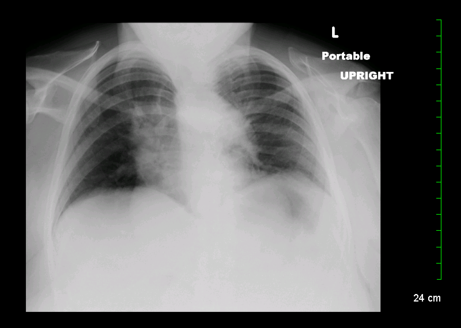
Posterior-to-anterior (PA): Exactly as it sounds, a PA view entails X-rays penetrating through the posterior/dorsal surface of a patient and being recorded on the opposite side. This is the preferred imaging orientation, however bed-ridden patients may not be able to allow for this type of X-ray to be taken.
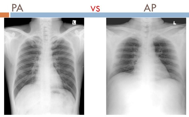
Anterior-to-posterior (AP): The opposite of a PA. this orientation is commonly taken with portable X-rays with patients who are bed-ridden and laying on their back. The anatomy of the heart can appear artificially larger due to this image orientation. In most circumstances PA orientations are preferred. It is important to realize that the LEFT side of the image (for both AP and PA films) represents the RIGHT side of the patient.
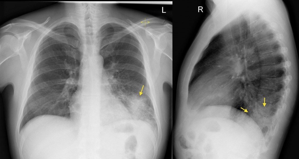
Lateral views (right/left): often, a lateral view usually accompanies a PA/AP chest X-ray. This can be helpful in settings where the single view is limited in localizing pathology (i.e. identifying the exact lobe of a lobar pneumonia in the right lung). The position of the spine on the lateral view can help inform is direction (if the image is taken from the right, the spine will be on the right side of the film and vice versa).
Left lateral decubitus position (LLDP): sometimes patients are radiographed when laying on their left side. This can be done for logistical reasons (patient is unable to stand for an upright lateral X-ray) or can be done to evaluate for the effect of gravity on pathological findings (i.e. to assess for layering of a pleural effusion).
QUALITY MEASURES FOR A CHEST X-RAY
Now that we are more familiar with X-ray machines, and common chest X-ray orientations, it is time for us to assess the quality of the X-ray image we are viewing. THIS SHOUDL BE DONE BEFORE FURTHER ANALYSIS!!! If an image is poor quality we can not use it for accurate diagnosis. Here are the 5 major considerations to make to evaluate if a chest X-ray is technically adequate.
- Penetration
- Inspiration
- Rotation
- Magnification
- Angulation
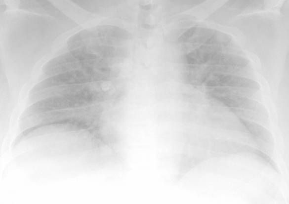
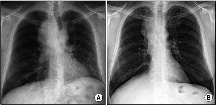
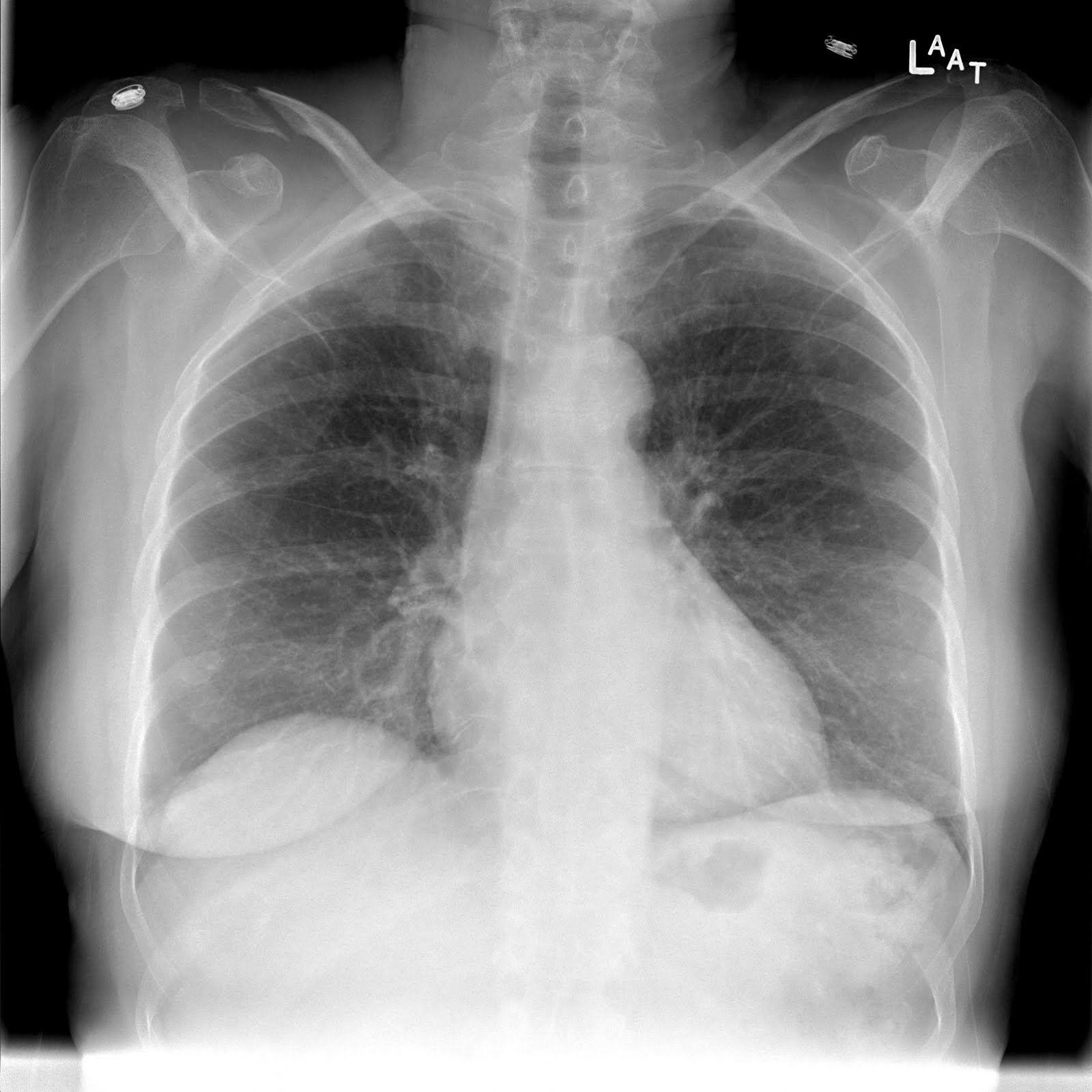
Penetration: Is the film under-penetrated? Penetration refers to how well the X-rays have penetrated the body of the patient. If film is under penetrated then there will be an excess of white present. One can tell if the film is under penetrated if the thoracic spine can not be seen through the heart.
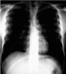
Penetration: Is the film over-penetrated? Alternatively, if the X-rays over penetrate the patient, there will be an excess of black present on the film. An over penetrated film will have lung markings that are decreased or absent.
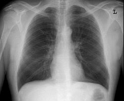
Inspiration: Has the patient inspired appropriately? In order to appreciate the anatomy within the chest cavity, it must be filled with air by having the patient inspire. On a PA view, 9 or more ribs should be visible and indicate proper inspiration by the patient.
Rotation: Is the patient centered? All too often patients are rotated when X-ray images are taken (especially on portable X-rays with patients lying askew in bed). This may not seem to be an issue, however it will complete distort the anatomy of the patient. The side rotated toward the X-ray machine will appear larger, and the side rotated away smaller. This can make structures (like the heart) appear larger/smaller then they really are. Look to the clavicles, length of the ribs, and position of vertebrae to help assess the positioning of the patient.
Magnification: How far away is the structure from the film when the image was taken? This can be a difficult concept to retain, however the further away a structure is from the X-ray film, the more it will MAGNIFY as the X-ray film is developed. It is for this reason that a standard PA (with the heart closer to the film) shows a more accurate size of the heart while an AP film (which increases the distance between the film and the heart) will show enlarged cardiac structures.

Angulation: Is the patient perfectly perpendicular when the image was taken? Similar to the concept of rotation, if the patient is not perpendicular to the X-ray machine taking the image, the view of the chest will become distorted. This is of more concern when taking X-ray images of supine patients, and can make the film difficult to interpret.
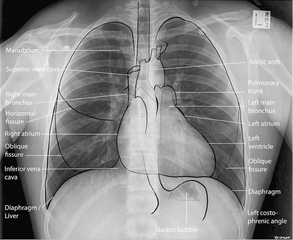
ANATOMY ON CHEST X-RAYS
With our quality measures out of the way, we can now go over what anatomy is possible to observe on a chest X-ray.
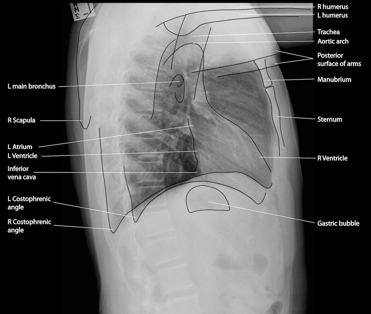
Let us not of course forget the lateral view as well.
As a quick reference click on the links below to learn more about radiological anatomy as they relate to the below topics:
SYSTEMATIC APPROACH TO INTERPRETING A FRONTAL CHEST X-RAY: ABCDE APROACH
As is the case with most things, reading X-rays benefits from having an intentional systematic approach. While not the only way, using the ABCDE acronym is helpful (in the setting of reading a PA or AP film):
- A (airway): looking at the trachea/airway is a good first step.
- B (bones): fractures can be overlooked if careful examination of the bones are not conducted
- C (cardiac): the heart (and associated structures) should be evaluated
- D (diaphragm): evaluating the diaphragms is a key step and can help contextualize pathology.
- E (everything else): a lazy category, this includes things like the drains and the lungs. It is typically a good idea to save the lungs for last, simply because their pathology seems to eclipse other findings.
FRONTAL VIEW: AIRWAY
While a simple starting point, evaluating the airway on a chest X-ray can help you orient yourself. Deviation of the airway can be easily overlooked and warrants investigation if detected.
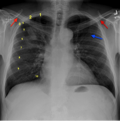
FRONTAL VIEW: BONES
An X-ray easily visualizes bones simply because of their increased density. It is a good idea to have a good systematic approach to evaluating bones on a chest X-ray (in order to make sure nothing is missed.
Clavicle (and bones of the upper body): the clavicles, humerus, and other bones of the upper body can be visualized in most chest X-rays. These can be quickly assessed for any obvious breaks/fractures.
Ribs: given proper penetration of the film, each of the ribs should be clearly visualized and should be assessed for fractures.
FRONTAL VIEW: CARDIAC
While perhaps not intuitive, a significant amount of information can be gained about the heart (and associated structures) from a chest X-ray. Here are some components to appreciate.
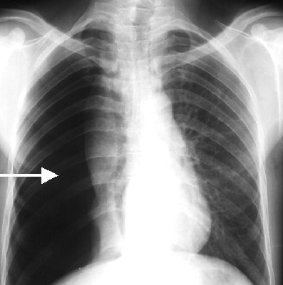
Mediastinum: the mediastinum is the central compartment of the chest cavity that houses the heart and its associated vessels. While other structures are also housed in this compartment (such as the trachea and esophagus), widening of this cavity can be suggestive of processes such as thoracic aortic aneurysm.
One of the initial evaluations can be to check the size of the heart. The cardiac width should be ≤ 50% of the thoracic width.

FRONTAL VIEW: DIAPHRAM
The nature of the diaphragm can provide some important context when reading a chest X-ray. Given the position of the liver, the right hemidiaphragm usually is situated at a higher level then the left hemidiaphragm. Some possible findings can include:
Flattened diaphragm: if the diaphragm is flattened in nature, this can suggest hyperinflation of the lungs (in obstructive diseases such as COPD).
Pneumoperitoneum is an important X-ray finding that can be indications of dangerous conditions (such as bowel perforation).
FRONTAL VIEW: EVERYTHING ELSE
A very “inclusive” section, there are many elements of the X-ray that have not been discussed yet. An important component that we have yet to discuss are the lungs. Many different lung findings can be found on a chest X-ray!
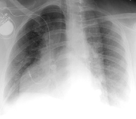
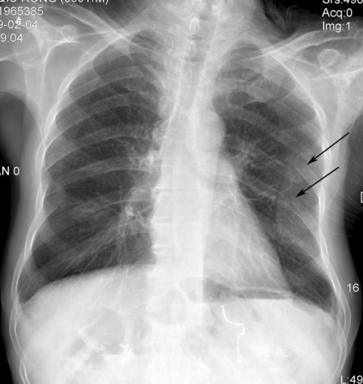
Pneumothorax: collapsed lungs can be observed due to the absence of lung markings in regions of the lung fields.
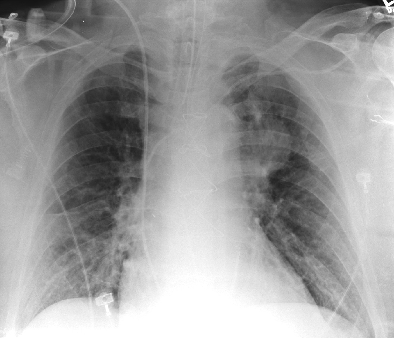
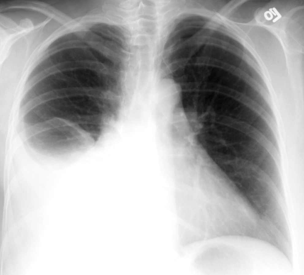
Pneumonia: a very common lung finding, X-rays can be used to definitively diagnose pneumonias.
- Right middle lobe pneumonias typically obscure the right side of the cardiac border
- Right lower lobe pneumonias typically obscure the right hemidiaphragm borders
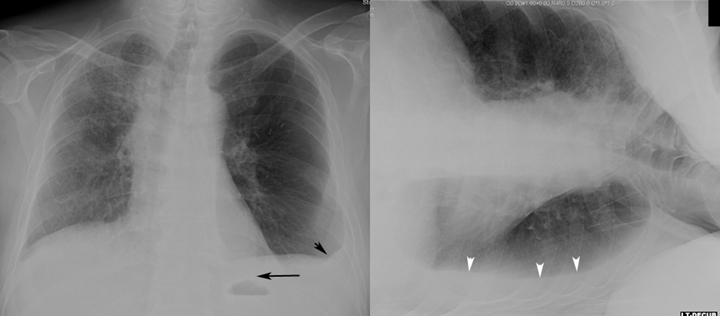
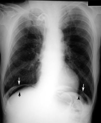
Pleural Effusion: collections of fluid in the pleural space can also be clearly visualized on chest X-rays.
SYSTEMATIC APPROACH TO INTERPRETING A LATERAL CHEST X-RAY
Lateral chest X-rays can often be ignored, however they have much utility! Here are 5 areas on a lateral chest X-ray to take a look at.
- Retrosternal clear space
- Hilar region
- Fissures
- Thoracic spine
- Diaphragm and posterior costophrenic sulci
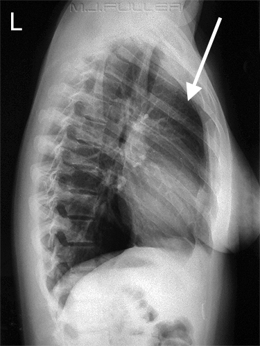
LATERAL VIEW: RETROSTERNAL CLEAR SPACE
The retrosternal space refers to the area right behind the sternum. Normally, there is a relatively clear crescent just behind the sternum and anterior to the shadow of the ascending aorta. If a mass is present here, it can suggest the presence of underlying pathology. It is important to keep in mind that if the patient can not lift there arms (the humorous/bones of the arm will be present in the X-ray view) this will “fill in” this normally clear space.
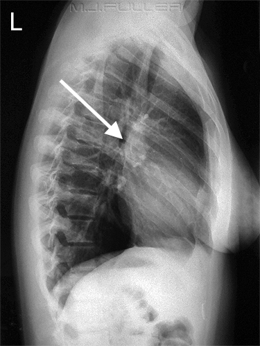
LATERAL VIEW: HILAR REGION
The hilar region refers to the “lung roots” that are found on the medial aspect of each lung. These areas can be sometimes difficult to evaluate on a frontal view. Much of the hilar region is made up of the pulmonary arteries. Normally there should be no mass in this area.
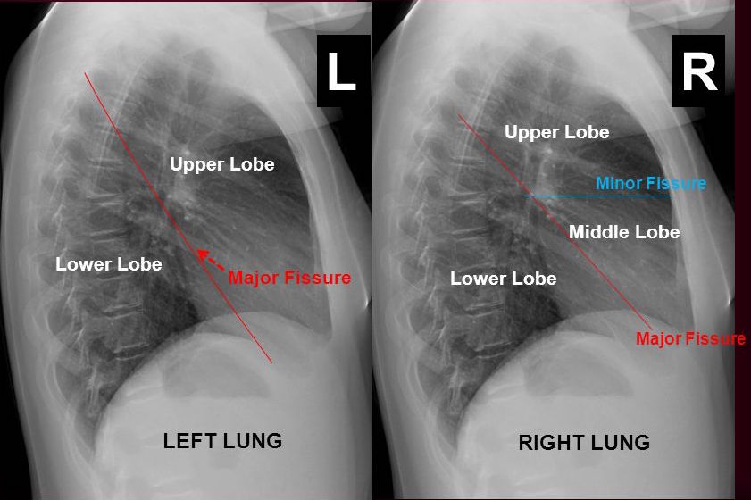
LATERAL VIEW: FISSURES
Lung fissures are the divisions between lobes of the lungs. These can be more easily appreciated on a lateral film as fine, white lines. Knowing the location of these fissures can help localize an opacity to a lung lobe. Whats more, thickening of these fissures can suggest the presence of fluid in the chest.
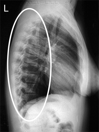
LATERAL VIEW: THORACIC SPINE
The thoracic spine can also be visualized on a later chest X-ray. Narrowing of the disk spaces and osteophytes can be observed. Changes in vertebral body height can also suggest the presence off compression fractures.
LATERAL VIEW: DIAPHRAM AND POSTERIOR COSTOPHRENIC SULCI
The diaphragm and the posterior costophrenic sulci can be visualized on a lateral chest X-ray. Blunting of the posterior costophrenic sulci can suggest the presence of a disease process such as a pleural effusion.

Page Updated: 10.07.2016