Page Contents
OVERVIEW
This page is dedicated to covering how the condition pleural effusion will appear on different types of radiological imaging studies.
BASIC CHARACTERISTICS OF A PLEURAL EFFUSION
Fundamentally a pleural effusion refers to the collection of fluid between the parietal and visceral pleura. There can be many different causes of this fluid collection (infection, malignancy, etc) however the cause of the effusion may not necessarily always be clear radiographically.
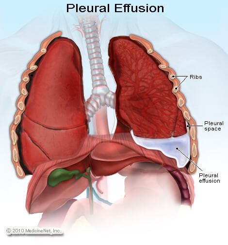
Here are some features of pleural effusion that can be observed across all imaging studies:
Signal intensity: given that three is fluid present here in this pleural space (instead of an air filled lung) the signal will be increased on most all imagine studies (increased white on a X-ray or CT scan).
Location: the pleural effusion will be observed in the pleural space (in the area of the lung fields) given its nature.
Gravity dependency: this fluid will observe the laws of gravity. In any study where the patient is upright, the fluid will collect at the base of the lungs. In any study where the patient is laying down (on their back or on their side) the fluid will collect at the lowest point closest to the ground.
There are a few different categories of pleural effusion that can be seen radiographically: It is useful in separating out pleural effusions by their relative size, simply because they will appear differently radiographically based upon the categories below:
- Minor pleural effusion: this refers to a very small pleural effusion that may not be visible on a frontal view (but may be seen on a lateral or LLDP).
- Moderate pleural effusions: this is a very generic “catch all category”. Basically any pleural effusion that does not fall into the other two categories is a moderate (or just a regular) pleural effusion.
- Massive pleural effusions: this is a very large pleural effusion that involves the entire lung. It will cause an entire lung “white out” which is referred to as a hemithorax opacification.
CHEST X-RAY
A chest X-ray can be a very common study by which a pleural effusion will be diagnosed. Make sure to review how to read chest X-rays, as well as the archive of unremarkable chest X-rays as a reference point.
Some key features to keep in mind for the appearance of pleural effusions on a chest X-ray are:
- Presence of opacity: fundamentally the fluid that comprises the pleural effusion will appear as an opacity over a portion of the lung field. On a chest X-ray this will show up as “increased white”.
- Air/fluid level or fluid meniscus: given that a pleural effusion is a collection of fluid, it is very common for this condition to have a clear air/fluid level visualized radiologically.
- Mediastinal shift AWAY from opacification: in certain cases the collection of fluid in a pleural effusion will cause the shift of mediastinal structures away from the effusion (due to mass effect)
MINOR PLEURAL EFFUSIONS
The archive below organizes different examples of a minor pleural effusions. These effusions often are diagnosed with the utilization of more then one view (there will be fluid “layering” for example on a LLDP view). Click on the thumbnails below to view the archive.
MODERATE PLEURAL EFFUSIONS
The archive below organizes different examples of a moderate pleural effusions. These are the “classic” pleural effusions that are seen. Click on the thumbnails below to view the archive.
MAJOR PLEURAL EFFUSIONS
This archive below organizes different examples of a massive pleural effusion. This will result in a complete white out of the affected lung. Make sure to review the page discussing this radiological finding of complete lung “white out” called hemithorax opacification. Click on the thumbnails below to view the archive.
COMPUTIRIZED TOMOGRAPHY (CT-SCAN)
A pleural effusion can also be visualized on a CT scan, and given how common CT scans are becoming, it is useful to understand how a pleural effusion will appear on this imaging modality.
Some key features to keep in mind for the appearance of pleural effusions on a CT-scan are:
- Presence of signal: fundamentally the fluid that comprises the pleural effusion will appear as an opacity over a portion of the lung field. On a CT- scan this will show up as an increased signal (often grey).
- Air/fluid level or fluid meniscus: given that a pleural effusion is a collection of fluid, it is very common for this condition to have a clear air/fluid level visualized radiologically.
- Gravity dependent nature: the fluid of a pleural effusion will collect posteriorly because a patient is on their back for a CT-scan.
Here is an archive of how pleural effusions will appear on a CT-scan: These are all put in one large archive for now. Click on the thumbnails below to view the archive.
MAGNETIC REASONANCE IMAGING (MRI)
A pleural effusion can also be visualized on a MRI scan
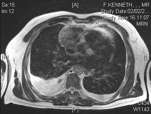
Some key features to keep in mind for the appearance of pleural effusions on an MRI are:
- Presence of signal: fundamentally the fluid that comprises the pleural effusion will appear as an opacity over a portion of the lung field. On an MRI this will show up as an increased signal (color will depend on the type of MRI).
- Air/fluid level or fluid meniscus: given that a pleural effusion is a collection of fluid, it is very common for this condition to have a clear air/fluid level visualized radiologically.
- Gravity dependent nature: the fluid of a pleural effusion will collect posteriorly because a patient is on their back for the MRI.
Page Updated: 10.11.2016