This page is dedicated to covering how the trachea will appear across different radiological studies.
CHEST X-RAY
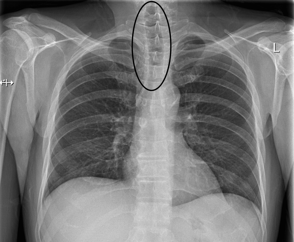
A chest X-ray is a very routinely ordered study, and a core component of reading a chest X-ray is to evaluate the appearance of the trachea/airway.
The trachea (circled) appears slightly darker then surrounding structures because it is air filled. X-rays will have an easier time penetrating this structure to develop the film behind the patient (source)
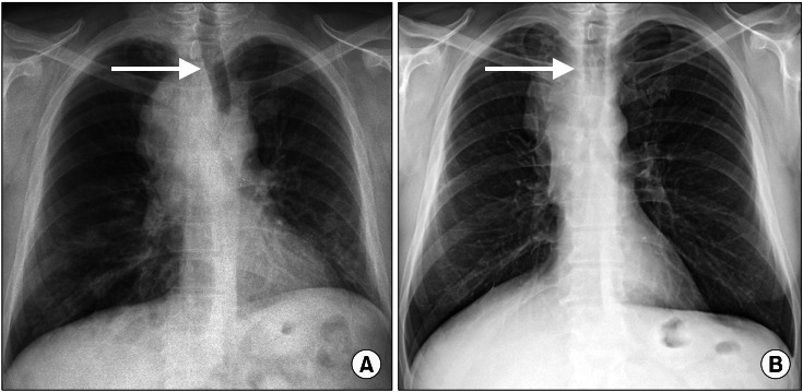
Deviation of the trachea/airway is an important characteristic to keep an eye out for
In pane A a large mass around the trachea has caused significant deviation. In page B this issue has been resolved and the trachea is midline (source)