Page Contents
OVERVIEW
The following guide helps walk you through a comprehensive approach to understand and interpreting a non-contrast head CT scan. As a refresher read this guide on the fundamentals of CT studies to help orient yourself.
WHEN IS IT ORDERED?
It is important to appreciate when a non-contrast head CT scan might be ordered.
To evaluate for acute intracranial bleeding: a non-contrast head CT is the preferred study that is ordered to evaluate for the presence of any acute bleeding in the cranium.
In tandem with a IV contrast head CT: sometimes head CT scans with and without IV contrast are ordered together. This can be done to evaluate how the addition of IV contrast alters pathology that is visualized on the scan.
SIGNAL INTENSITIES ON A HEAD CT SCAN
It is important to understand what types of signal densities may represent on a head CT scan.
Hypodense/Hypointense Signal (Dark)
- Fat: not usually present IN the cranium
- Air: such as the air in the sinuses
- Water: such as the CSF found in the ventricles
- Old blood: such as a chronic subdural hematoma
Isodense Signal
- Normal brain: white matter appears darker then grey matter due it its increased fatty content (myelin)
- Some forms of protein: subacute subdural hematoma
Hyperdense/Hyperintense Signal (Light)
- Metal: such as aneurysm clips or bullets
- Iodine: this is present in the IV contrast (NOT present in a non-contrast head CT scan).
- Calcium: such as bone or calcified pathological findings
- Hemorrhage: primarily acute hemorrhage (with its high protein content) is very dense.
ORIENTATIONS OF THE HEAD CT SCAN
There are few important orientations that can be used to view a head CT scan that are worth reviewing here.
Axial Orientation: This orientation shows images that are parallel to the top of the head.
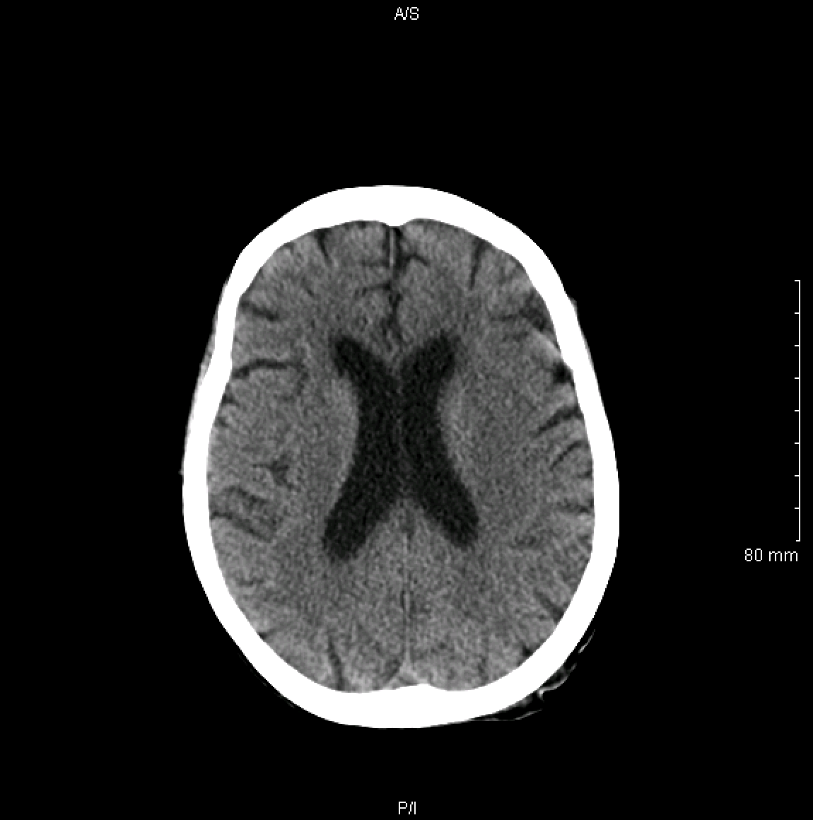
When reading an axial image radiographically it is important to remember a few things:
- The top of the image represents the anterior surface of the patient.
- The bottom of the image represents the posterior surface of the patient.
- The right side of the image represents the left side of the patient.
- The left side of the image represents the right side of the patient.
- When scrolling through the image one travels either inferiorly or superiorly through the structure being analyzed
Coronal Orientation:
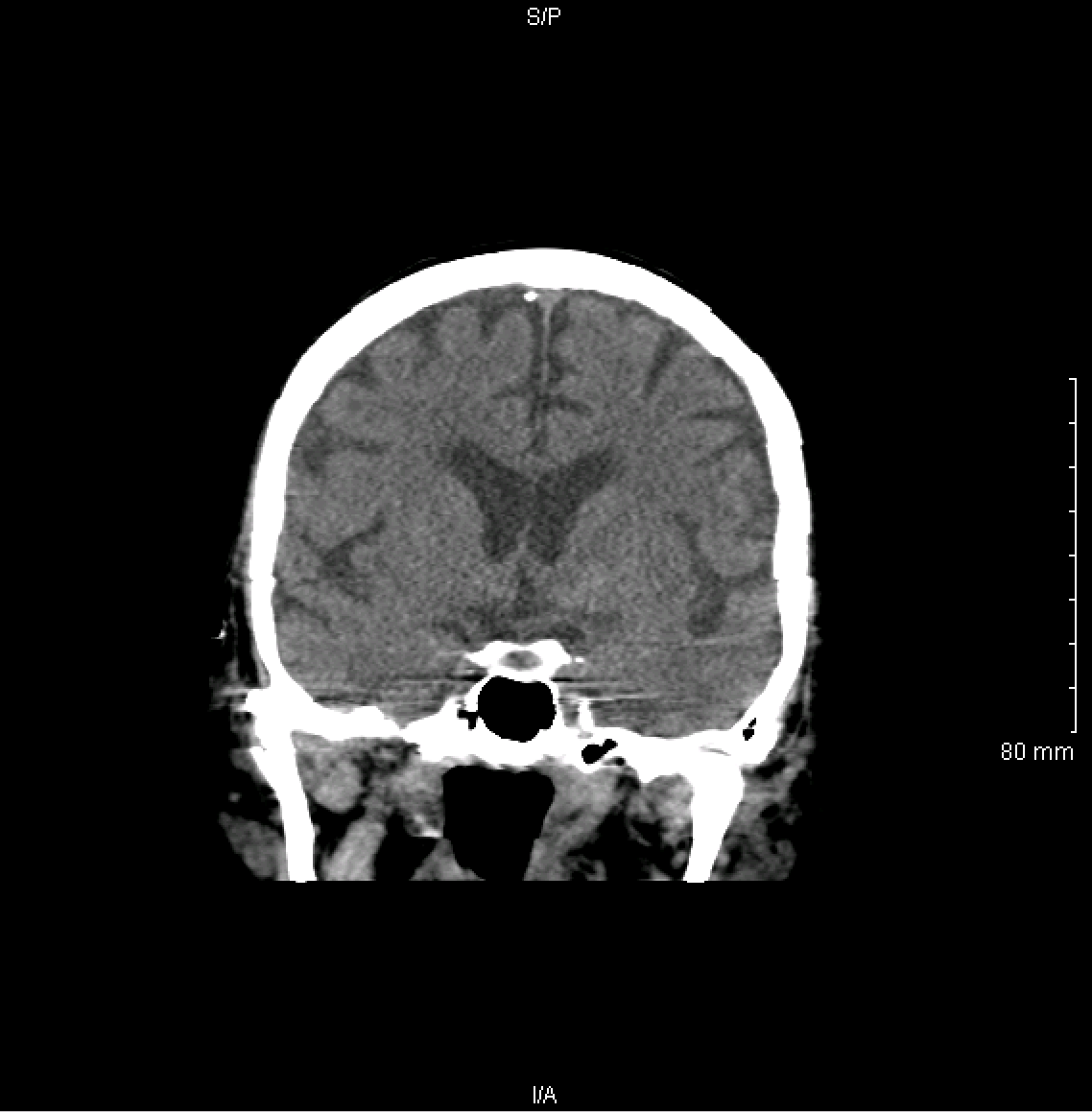
When reading a coronal image radiographically it is important to remember a few things:
- The top of the image represents the superior surface of the patient.
- The bottom of the image represents the inferior surface of the patient.
- The right side of the image represents the left side of the patient.
- The left side of the image represents the right side of the patient.
- When scrolling through the image one travels either anteriorly or posteriorly through the structure being analyzed
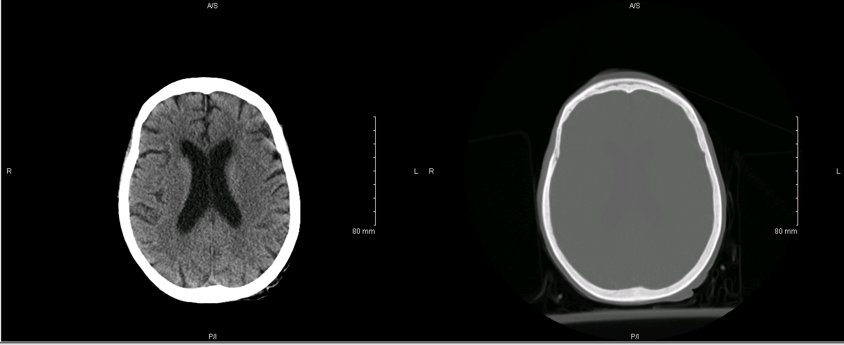
DIFFERENT “WINDOWS” ON THE HEAD CT SCAN
Without getting too complicated, it is important to realize that on a traditional head CT scan there are different “windows” that can be viewed. Typically there is a brain window and bone window that helps to highlight the brain and bone respectively (for improved analysis).
ANATOMY ON THE HEAD CT SCAN
While it is difficult/impossible to summarize all of the relevant anatomy in a single set of images the below links help to highlight how some important intracranial structures are visualized radiologically.
- Portions of the brain: cerebellum
- Ventricles of the brain: lateral ventricles
APPROACH TO READING A NON-CONTRAST HEAD CT SCAN
When reading a non-contrast head CT scan a systematic approach can be used to ensure that nothing is missed. There are a few categories we can analyze separately.
- Bones: the skull can be evaluated for fracture
- Bleeding: the non-contrast head CT is the study of choice for detecting acute bleeding.
- Brain pathology: masses, calcifications, loss of gray/white matter differentiation, and any issues in the brain parenchyma should be evaluated.
- Ventricles: abnormally sized ventricles should be assessed when reading a head CT scan.
ALGORITHM
Below is a general approach on how to read a head CT-scan
- Soft tissues: begin using a soft tissue window and look around the skull for any signs of trauma or contusion. This can be done in all 3 of the views.
- Bones: use the bone window and look for any fractures
- Skull
- Nasal bones
- TMJs
- Sinuses/air spaces: continue on the bone window and look at all the sunuses and the mastoid air cells as well.
- Brain parenchyma: use the brain windows and look for any pathology. There are specific places to look.
- Insular cortext
- Basal ganglia (globus pallidus/Putamen)
- Ventricles: any hydrocephalus?
Page updated: 10.10.2016