OVERVIEW
This page is dedicated to covering the fundamental aspects of computed tomography (CT) imaging. Given its similarities, it may be useful to first cover the fundamentals of X-ray studies page before reading this guide.
How It Works:
Considerations Before The Scan:
Considerations During The Scan:
- Where to image
Considerations After The Scan:
- Reformatting the CT-scan
- Slice thickness
- Algorithms
- Windowing
SETTINGS USED IN CT SCANS
Timing of image aquisition (after giving IV contrast)
Slice thickness
HOUNDSFIELD UNITS
Houndsfield units (HU) are the units used to measure the density of matter on a CT-study: the higher the HU value the more dense an object is. Here are some important values to keep in mind on the HU scale.
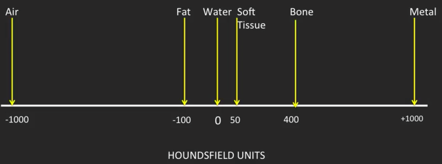
Other values include:
- Clotted blood: ~ 70 HU
WINDOWS
While the HU scale ranges from -1000 to +1000 there are limited shades of grey that can be utilized by radiological software/can be detected by the human eye. It is for this reason that different “windows” exist that can be used to modify CT studies.
A window essentially refers to a specific dedicated range of HU in which different densities can be distinguished by various shades of grey. Anything less dense then the range of the window will appear as black on the scan, and anything more dense will appear as white. The narrower the window the more resolution in differentiating structures with slight differences in density. Examples of different windows include:
- Abdominal window
- Bone window
- Brain window
- Liver window
- Lung window
- Mediastinum window
Page Updated: 09.05.2016