Page Contents
OVERVIEW
This page is dedicated to covering the fundamental aspects of how ultrasound imaging works.

WHAT ARE THE BASIC COMPONENTS OF AN ULTRASOUND MACHINE
While there are may different types of ultrasound machines, they all (more or less) share certain common shared elements. Lets take a moment to cover these now:
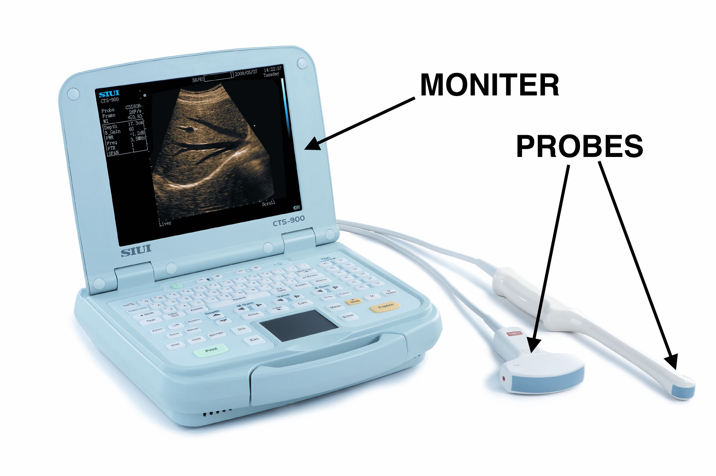
The probe is responsible for generating AND detecting sound waves.
The monitor is responsible for rendering an image of the structures that are being “probed”.
HOW DOES IT WORK?
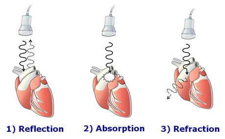
Fundamentally ultrasound imaging works by utilizing sounds waves to visualize structures within the body. A transducer (i.e. the ultrasound probe) will emit sound waves, and will also record echoing waves that bounce back towards it off of structures. The image that is generated is combination of sound waves that are reflected, absorbed, and refracted.
In looking at a real ultrasound image it is important to appreciate exactly how the detection of sound waves translates into the generated picture that is visualized. The probe that is used for ultrasound can detect the amount of signal that is reflected from internal structures (which can evaluate the composition of the object), as well as the amount of time that it takes for these waves to be reflected (which can assess for the distance of the object in question).
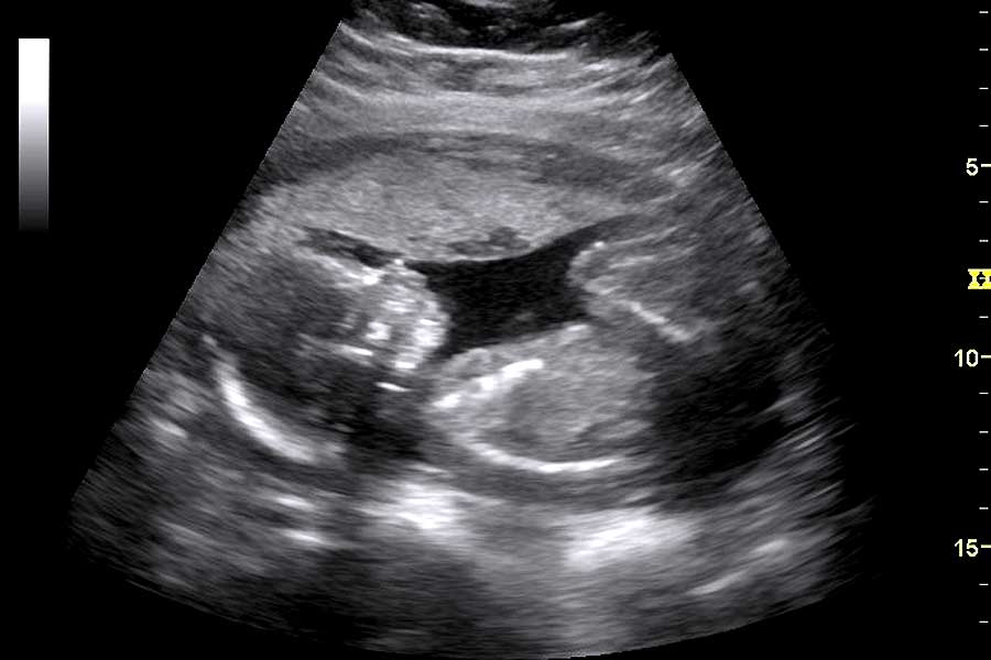
Here are a few basic components to keep in mind when looking at an ultrasound image:
- Lighter colors (white/echogenic/hyperechoic) represent structures that have reflected the produced sound waves back to the probe (the more a structure reflects sound waves the lighter it will appear in the image). Bones will reflect sound waves well and appear white.
- Darker colors (black/hypoechoic/anechoic) represent structures that have absorbed/do not reflect the produced sound waves (the less a structure reflects sound waves the darker it will appear on the image). Fluid does not reflect sound waves and will appear black.
- The top of the screen (anterior) shows structures that are closer to the probe (these are areas in which it takes less time for sound waves to reflect back to the probe).
- The bottom of the screen (posterior) shows structures that are further away from the probe (these are areas that take the most time for the sound waves to reflect back to the probe)
WHAT DOES DOPPLER ULTRASOUND REFER TO?
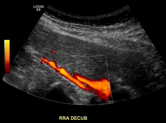
Without getting into too much detail, the idea of Doppler ultrasound is actually (at its core) quite simple. Given the nature of the doppler effect, doppler ultrasound is useful because it can actually determine the speed at which objects that are being evaluated are moving.

Why do we care about doppler ultrasound in medicine? Really in the realm of the medical field doppler ultrasound becomes very useful went evaluating for the presence of blood flow (given that pumping blood in arteries really is what moves the most in the body). Doppler ultrasound can be combined with traditional ultrasound imaging (this is often called Duplex ultrasonography) to provide information on both structure and blood flow within areas of the body.
OPTIMIZING YOUR UTILIZATION OF ULTRASOUND
This guide was created to go into further detail about how to adjust ultrasound machine settings to that the user can optimize the quality of images that they can acquire from this modality.
Page Updated: 08.15.2016