Page Contents
OVERVIEW
This page organizes relevant information, anatomy, and pathology relating to the third cranial nerve (the oculomotor nerve).
FUNCTION
The third cranial nerve controls eye movements which are depicted in the figure below (which shows the views of the clinical facing the patient and performing a routine eye exam where the eyes move in the in the charactersitc “H” pattern).
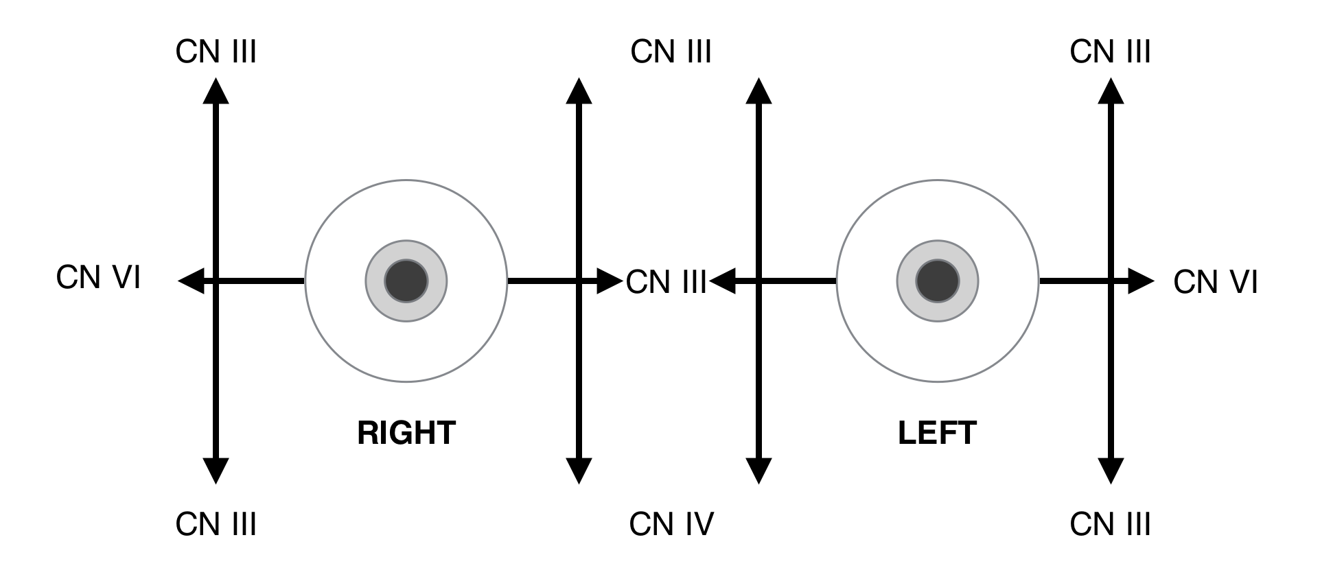
**CN III innervates every muscle except for the Superior Oblique (CN IV) and the Lateral Rectus (CN VI), additionally it will control elevation of the eyelid and damage to the nerve causes dilation given its innervation from the Edinger-Westphal nucleus.
ORIGIN: CRANIAL NERVE 3 BRAINSTEM NUCLEI
The third cranial nerve begins first in the midbrain, where it is supplied by two nuclei:
- The oculomotor nucleus: this supplies somatic efferent fibers to the oculomotor nerve. It is located anterior/ventral to the periaqueductal grey matter in the midbrain at the level of the superior colliculus. It is just inferior and dorsal to the level of the red nuclei (depending on the orientation of an axial slice).
- The Edinger-Westphal nucleus: supplies parasympathetic fibers to the oculomotor nerve which synapse at the ciliary ganglion (it is immediately behind/posterior to the oculomotor nucleus at the same level in the midbrain).
To locate the general region of these nuclei, it is easiest to find the midbrain, look closely at the cerebral aqueduct, and scroll to the level of the superior colliculus (dorsal bumps will be appreciated along the midbrain). At this level the nuclei will be just anterior to the periaqueductal grey matter (on either medial side of the brainstem). This is depicted below.

** Notably the fibers to the inferior rectus, inferior oblique, and medial rectus muscles supply the ipsilateral eye; fibers innervating the superior rectus muscle decussate and supply the contralateral eye.
**The decussating fibers pass through the opposite superior rectus nucleus; thus damage to the right oculomotor nucleus might have bilateral superior rectus muscle involvement
BRAINSTEM COURSE
After receiving supply from both nuclei, the nerve courses anteriorly through the brainstem with fibers crossing through the red nucleus, and through the medial aspect of the substantia niagra before exiting the midbrain along the medial aspect of the cerebral peduncle.
CISTERNAL COURSE
After exiting along the medial aspect of the cerebral peduncle, the oculomotor nerve will travel through the interpeduncular cistern, and then along the lateral aspect of the suprasellar/chiasmatic cistern before piercing the dura through the occulomotor cistern to enter the cavernous sinus.

CAVERNOUS COURSE