Page Contents
OVERVIEW
Cerebral angiographic projections have a direct influence on visualization of key vascular anatomy during procedures. This page covers some common projections (and how they change the appearance of key vascular anatomy).
THE RELATIONSHIP BETWEEN PROJECTIONS AND VASCULAR APPEARANCE
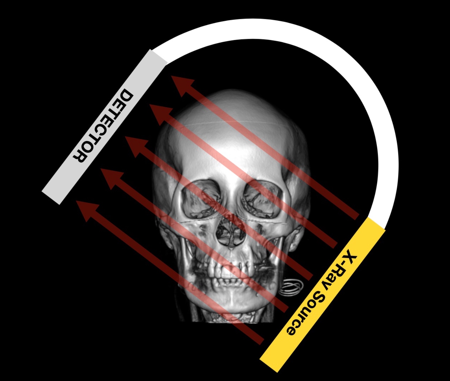
As the C arm is moved around, the path of X-ray beams will move with it. A projection will ELONGATE a vessel (improving its visualization) if the path of X-ray beams are perpendicular to that vessel. Contrastingly a projection will SHORTEN a vessel (decreasing its visibility) if the X-ray beams are parallel to that vessel.
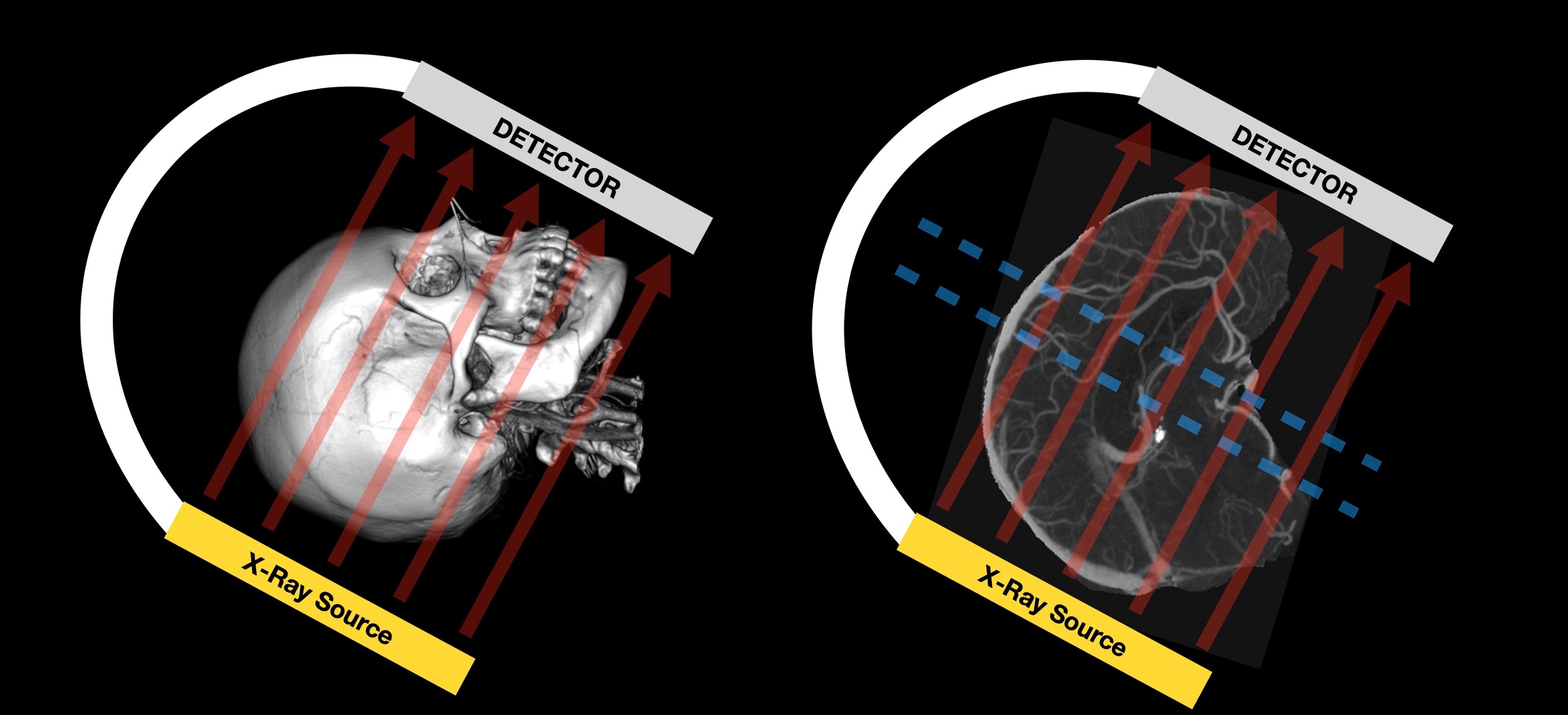
In summary the angle of x-ray beams relative to a vessel changes its appearance: if the angle is perpendicular the vessel is ELONGATED, if the angle is parallel the vessel is SHORTENED.
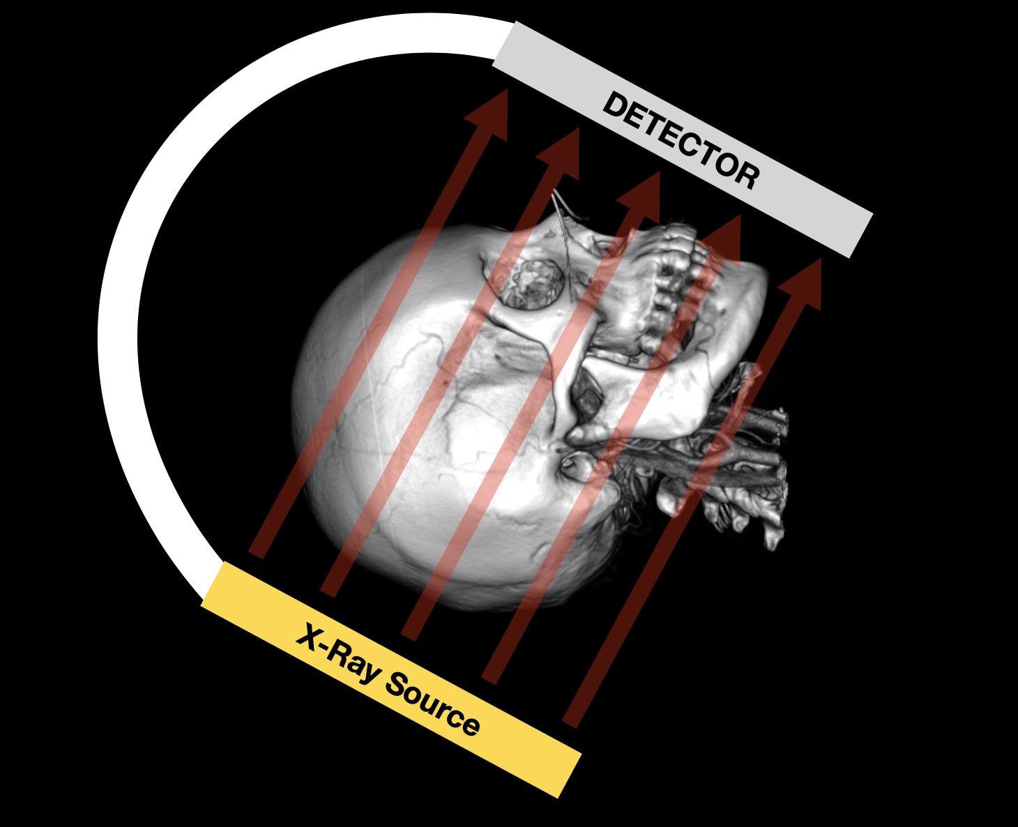
TOWNE’S VIEW
The Townes projection is a common frontal projection: The detector is angled up toward the vertex of the skull.
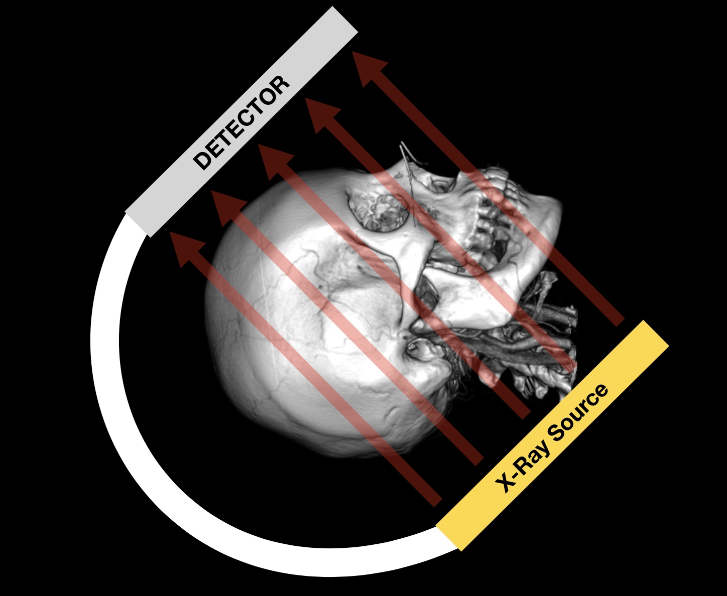
Here are the key features:
- Should see the IACs in the petrous bone but don’t move indicator towards the point of seeing the foramen magnum.
- This puts the X-ray beam orthogonal to the PCOMS and PCAs
- This ELONGATES the PCOMs and PCAs
- Will conversely shorten the basilar artery
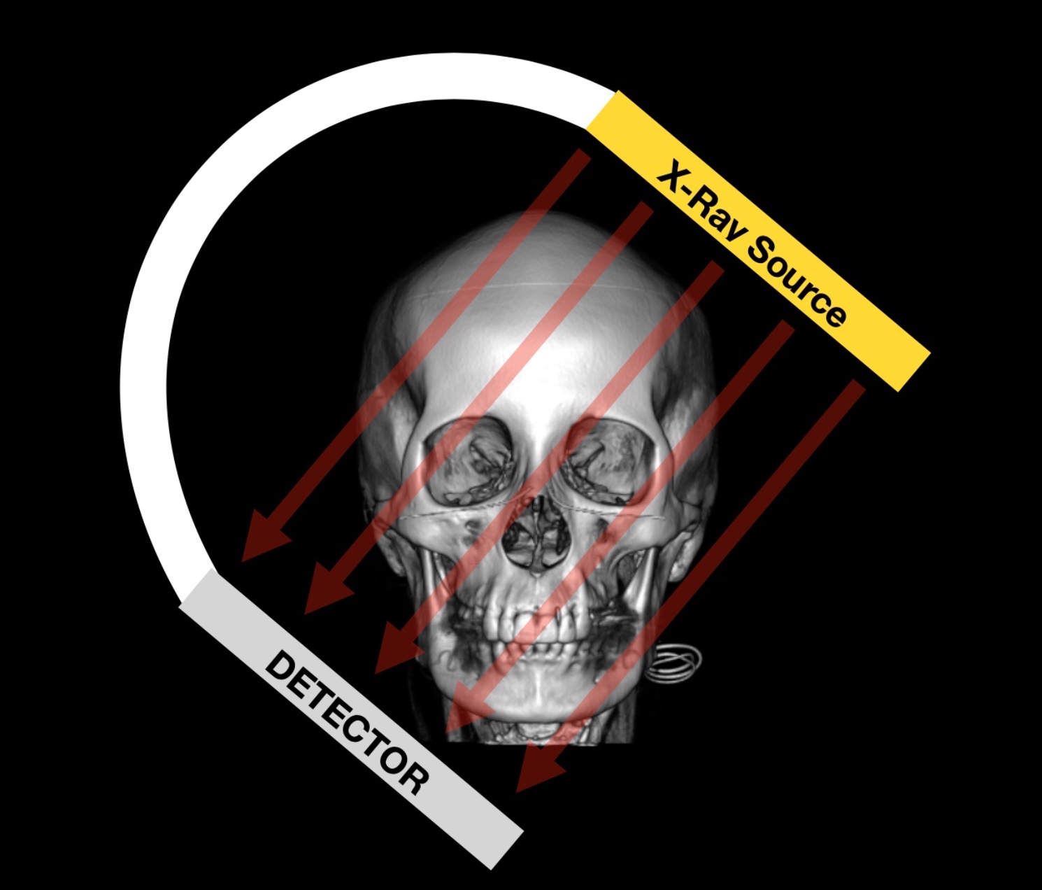
WATERS’ VIEW
The Water’s projection is a common frontal projection: The detector is angled down toward the patient’s maxilla.
Here are the key features:
- Orbital roofs should be just off the top of the screen (depending on the mag).
- This puts the X-ray beam orthogonal to the basilar artery
- This ELONGATES the basilar artery as well as its branches
SCHULLER’S VIEW
The Schuller’s projection is a common lateral projection: Angle detector to the shoulder of the side your catheter is on.
Here are the key features:
- Elevates the M2, M3s, and M4s and separates them from the ACA.
- Shortens the M1 branch.
- Some PCA branches more visible
REVERSE SCHULLER’S VIEW
The reverse Schuller’s projection is a common lateral projection: Angle detector away from the shoulder of the side your catheter is on.
Here are the key features:
- Lowers the MCA branches.
- Puts the ACOM and A1 in plane.