Page Contents
OVERVIEW
Often times ultrasound will be utilized at some point in the peri-procedural setting for interventional radiology cases. This can include anything from scanning the patient prior to prepping them for the case, to performing parts of the case with ultrasound guidance. Ultrasound is a powerful tool, and this page is dedicated to covering the most critical aspects when it comes to utilizing ultrasound in the peri-procedural setting.
This topic has various subcomponents that have been organized into the various sections below.
PICKING THE RIGHT EQUIPMENT
It all begins with picking the correct equipment to utilize:
- Selecting the ultrasound machine
- Selecting the ultrasound probe(s)
POSITIONING AND POSTURE
Commonly overlooked, however proper positioning and posture is critical for success when utilizng ultrasound:
- Body posture and patient positioning
- How to hold the ultrasound probe
- Common scanning positions
OPTIMIZING ULTRASOUND MACHINE SETTINGS
Not all ultrasound machine settings are created equal, there are various adjustments that can be made to give the best image:
- Adjusting the frequency setting
- Adjusting the scanning depth
- Setting the focal zone
- Adjusting the gain setting
- Adjusting the time gain compensation setting
- Using the zoom feature
EVALUATING VESSELS/BLOOD FLOW
In addition to grayscale evaluation, it can be useful/necessary to evaluate vascular anatomy utilizing other ultrasound features:
- Utilizing color doppler
- Utilizing power doppler
- Utilizing B mode
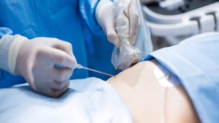
NEEDLE LOCALIZATION WITH ULTRASOUND
Ultrasound guided needle localization can be challenging given the dynamic nature of the task:
- How to hold the syringe/needle
- Positioning of the needle next to the probe (linear probe)
- Positioning of the needle next to the probe (curved probe)