OVERVIEW
This page aims to break down all of the basic components of an electrocardiogram (EKG/ECG) tracing. Each area of the tracing corresponds to a certain type of cardiac physiology, and understanding these relationships may help down the line with reading and interpreting these clinical studies.
BASIC COMPONENTS
An electrocardiogram (EKG or ECG for short depending on where you are from) in a nutshell is a electrical snapshot/image of the hearts activity from a particular vantage point (more on this later). Things can get complicated very quickly when trying to decipher EKGs, and the most important thing is to work through the information in a systematic process.
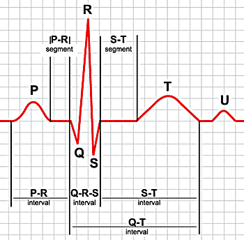
The first thing we must do is evaluate what we are hoping to actually see on the EKG. When looking at the typical image of a EKG (below) it is important to realize what each portion of the tracing represents physiologically.

P wave: is the first short upward movement of the EKG tracing. It indicates that the atria are contracting, pumping blood into the ventricles.
The QRS complex: normally beginning with a downward deflection (Q), a larger upwards deflection (R); and then a downwards S wave. The QRS complex represents ventricular depolarization and contraction.
T wave: is normally a modest upwards waveform representing ventricular repolarization.
U wave: is sometimes seen and marks re-polarization of the purkinje fibers.
Page Updated: 03.04.2018