OVERVIEW
The topic of percutaneous feeding tubes can be a complicated one given the amount of different types of tubes that are utilized clinically. While many different manufacturers and styles of tubes exist, there are core anatomical components that are present within most all percutaneous feeding tubes.
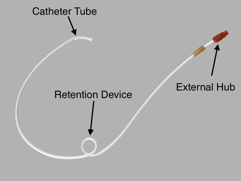
These core components of percutaneous feeding tubes include:
- External hub
- Catheter tube
- Retention device
EXTERNAL HUB
The external hub of the feeding tube is what will be externally visible during the physical exam. Often times this will be the first clue in identifying what type of tube a patient has. There are a few features of the external hub that are important to pay particular attention to:
- Number of ports: counting the number of ports a feeding tube’s hub has can reveal more about what exactly is present on the inside of the patient. Each lumen of a feeding tube will have its own port, and a balloon retention device (more on this below) will also have its own port as well. Keeping this in mind, when a tube hub is seen that only has 1 port (like the tube above) one already knows that there is only one lumen in the tube, and that there is no balloon retention device present.
- Profile of the hub: the degree of how flat a tube’s hub sits against the outer abdomen can also be important to appreciate. Some brands of tubes have special “low-profile” versions of their tubes.
The gallery below shows examples of the external hub from different percutaneous feeding tubes. Click on the thumbnails below to open up the gallery:


CATHETER TUBE
The actual tube of the feeding catheter is perhaps the largest anatomical structure present. The function of the percutaneous feeding tube in question is dictated by the features present/absent within this tube.
- Number of lumens: the number of lumens in the tube is often related to its function.
- Location of tip/tube holes: this is a feature of the tube that is closely related to the number of lumens that the tube contains. Major locations of the tube tip/tube holes will be the stomach and/or the jejunum.
The gallery below shows examples of catheter tubes from different percutaneous feeding tubes. Click on the thumbnails below to open up the gallery:

RETENTION DEVICE
Given that placed feeding tubes run the risk of becoming misplaced/falling out, a core anatomical feature of these catheters is that they will contain some type of retention device aimed at keeping the tube in place. There are a few types of retention devices that are commonly used.
- Balloon: an inflatable balloon is often used to keep either the tip or a portion of the feeding tube in place. It is important to keep in mind that the presence of an inflatable balloon will also be associated with an additional port on the external hub of the tube (that are used for inflating the balloon).
- Securing external disk: some brands of feeding tubes will have an external disk that is used to secure the outer portion of the tube, and can be slid towards the internal balloon to keep the whole feeding system in place.
- Pigtail/coil: Often times feeding catheters that don’t have a securing balloon will instead rely upon a pigtail/or coil in the tube to keep it in place. The shape of these tubes can be appreciated radiographically.
The gallery below shows examples of retention devices from different percutaneous feeding tubes. Click on the thumbnails below to open up the gallery:

Page Updated: 09.10.2017