Page Contents
OVERVIEW
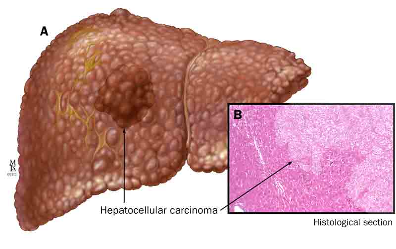
This page is dedicated to covering how the condition hepatocellular carcinoma (HCC) will appear on different types of imaging studies.
BASIC CHARACTERISTICS
Fundamentally, HCC is a malignancy that involves the liver.

Here are some general features of this condition that might be appreciated across modalities:
- Arterial enhancement
- Large size
- Rapid growth
ULTRASOUND
Focal liver nodules can be detected on abdominal ultrasound, however it is important to appreciate that HCC does not necessarily have a “characteristic” appearance on ultrasound (and can present variably on this imaging modality).
Key features of the appearance of this condition on this imaging modality are:
- Hypoechoic nature of lesions (typically but not always): in some cases might be hyperechoic or have mixed echogenecity
- Size larger then 1 cm: the majority of liver lesions that are less then 1cm in size at the time of detection are not HCC
- Enlarging nodules over serial ultrasound studies is more concerning for HCC
COMPUTERIZED TOMOGRAPHY (CT-SCAN)
Key features of the appearance of this condition on this imaging modality are:
MAGNETIC RESONANCE IMAGING (MRI)
Key features of the appearance of this condition on this imaging modality are:
FURTHER READING
Page Updated: 08.30.2017