Page Contents
- 1 OVERVIEW
- 2 BASIC CHARACTERISTICS
- 3 COMPUTERIZED TOMOGRAPHY: NON-CONTRAST HEAD CT-SCAN
- 4 COMPUTERIZED TOMOGRAPHY: CT ANGIOGRAPHY (CTA)
- 5 MAGNETIC RESONANCE IMAGING: SUSCEPTIBILITY SEQUENCE HEAD MRI (GRE/SWI)
- 6 MAGNETIC RESONANCE IMAGING: T1 WEIGHTED HEAD MRI
- 7 MAGNETIC RESONANCE IMAGING: T1 WEIGHTED HEAD MRI (WITH CONTRAST)
- 8 MAGNETIC RESONANCE IMAGING: T2 WEIGHTED HEAD MRI
- 9 MAGNETIC RESONANCE IMAGING: FLAIR SEQUENCE HEAD MRI
- 10 SUMMARY OF TAKEAWAY POINTS
- 11 ACKNOWLEDGEMENTS
OVERVIEW
This page is dedicated to covering how cerebral cavernous malformation will appear on different types of imaging studies. It is important to note that cerebral cavernous malformations also go by a few other names (such as a cavernoma, or cavernous hemangioma) however for the purposes of this page, this condition will be referred to as a cavernous malformation. The video below explains a bit more about this page and how to utilize the galleries below:
BASIC CHARACTERISTICS
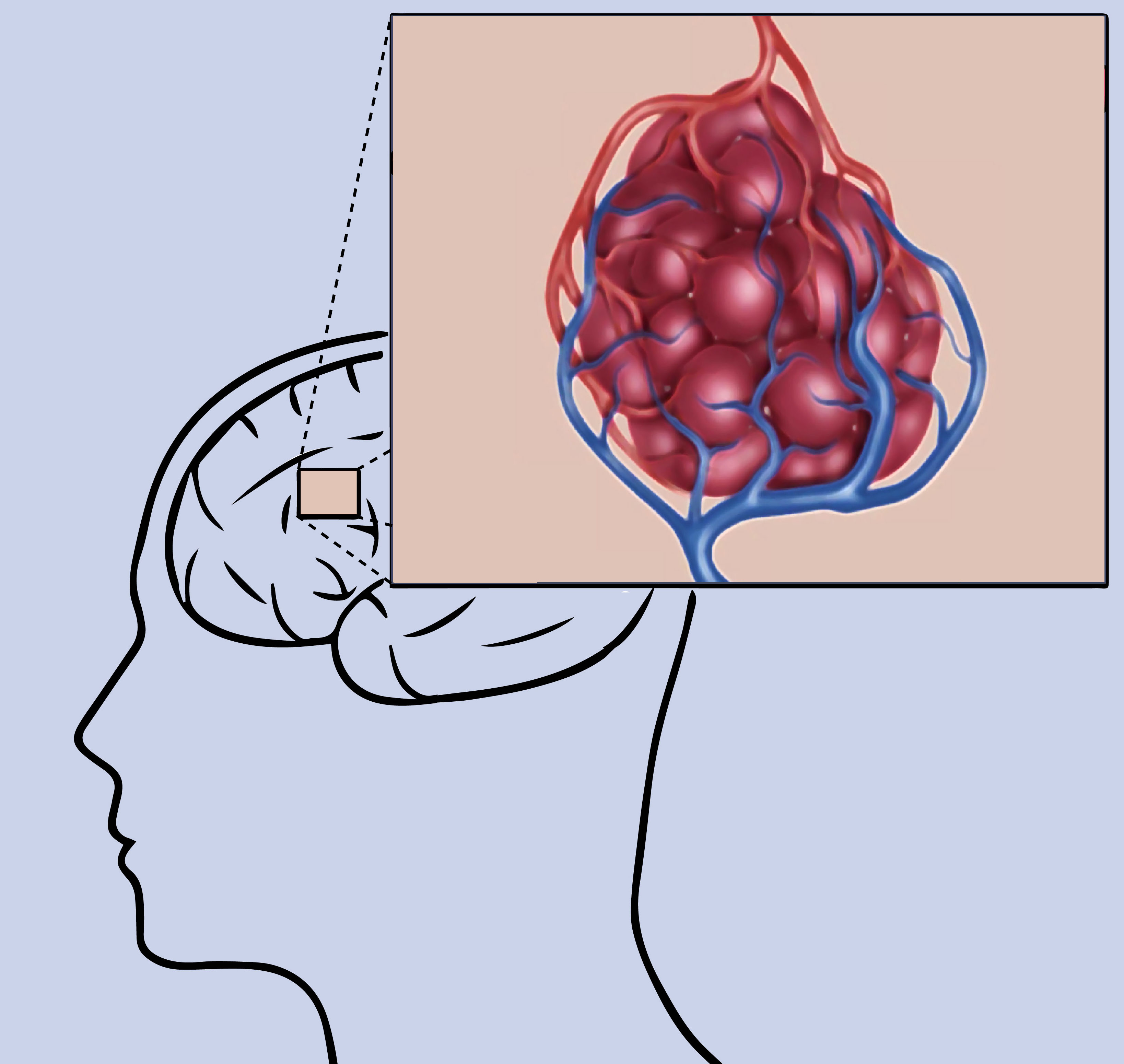
At their core, cavernous malformations are vascular lesions that are comprised of clusters of tightly packed, pathologically thin-walled small blood vessels (capillaries). They can displace normal neurological tissue depending upon their location. The vessels are filled with slow-moving or stagnant blood that is usually clotted or in a state of decomposition.
Here are some general radiological features of cerebral cavernous malformation:
- Clustered vessel appearance: given this condition’s nature, it can appear as a “mulberry-like” cluster or a “popcorn” lesion.
- Hemosiderin ring often surrounds the lesion and and can attenuate the signal from imaging modalities (such as causing a susceptibility artifact on MRI).
- Absence of normal brain parenchyma between interstices of these lesions: unlike a cerebra arteriovenous malformation there will be no normal brain tissue between the components of this lesion.
- Signs of hemorrhage/edema may be present if the lesion is/or has recently bled
COMPUTERIZED TOMOGRAPHY: NON-CONTRAST HEAD CT-SCAN
Key features of the appearance of cerebral cavernous malformation on this imaging modality are:
- Variable signal intensity: this condition may be hard to appreciate on this imaging modality, and often times may not show increased signal intensity. Larger/hemorrhaging lesions may demonstrate hypertensive signal however due to their increased density/presence of blood.
- Edema may be present if this lesion has recently bled.
Click on the thumbnails below to see examples of this condition utilizing this imaging modality:


COMPUTERIZED TOMOGRAPHY: CT ANGIOGRAPHY (CTA)
Key features of the appearance of cerebral cavernous malformation on this imaging modality are:
- Variable response to IV contrast: part of the time this lesion may enhance with the administration of contrast, however often it may not.
Click on the thumbnails below to see examples of this condition utilizing this imaging modality:


MAGNETIC RESONANCE IMAGING: SUSCEPTIBILITY SEQUENCE HEAD MRI (GRE/SWI)
Key features of the appearance of cerebral cavernous malformation on this imaging modality are:
- Dark signal on susceptibility sequences: a dark signal/absence of signal can be appreciated in the region of this lesion on susceptibility MRI sequences such as SWI and GRE. This may envelop the entire area of the lesion, or just a ring around it. It is the hemosiderin that surrounds this malformation that causes this artifact.
Click on the thumbnails below to see examples of this condition utilizing this imaging modality:


MAGNETIC RESONANCE IMAGING: T1 WEIGHTED HEAD MRI
Key features of the appearance of cerebral cavernous malformation on this imaging modality are:
- Variable signal intensity: this lesion has a variable signal intensity on T1 weighted images (sometimes it is bright, sometimes it is not)
- Classic “popcorn” or “berry” shape/appearance can sometimes be appreciated on T1 images.
- Dark ring artifact surrounding the lesion is sometimes seen on T1 weighted images
Click on the thumbnails below to see examples of this condition utilizing this imaging modality:


MAGNETIC RESONANCE IMAGING: T1 WEIGHTED HEAD MRI (WITH CONTRAST)
Key features of the appearance of cerebral cavernous malformation on this imaging modality are:
- Variable response to IV contrast: sometimes this lesion may enhance with the administration of contrast on T1, however often it may not.
Click on the thumbnails below to see examples of this condition utilizing this imaging modality:


MAGNETIC RESONANCE IMAGING: T2 WEIGHTED HEAD MRI
Key features of the appearance of cerebral cavernous malformation on this imaging modality are:
- Classic “popcorn” or “berry” shape/appearance can be best appreciated on T2 weighted images.
- T2 bright: the lesions are typically bright on T2 weighted imaging.
- Dark ring artifact surrounding the lesion is typically seen on T2 weighted imaging.
Click on the thumbnails below to see examples of this condition utilizing this imaging modality:


MAGNETIC RESONANCE IMAGING: FLAIR SEQUENCE HEAD MRI
Key features of the appearance of cerebral cavernous malformation on this imaging modality are:
- Appearance/features similar to T2 weighted images: these lesions look similar on FLAIR as they would on a standard T2 weighted images (popcorn lesion with surrounding dark ring)
- Signs of edema may be present if the lesion is hemorrhaging/or has bled recently (areas of FLAIR signal intensity may or may not be present).
Click on the thumbnails below to see examples of this condition utilizing this imaging modality:


SUMMARY OF TAKEAWAY POINTS
As a quick review here are the things to keep in mind about the radiological appearance of this condition:
- The lesion has a variable appearance on CT scans: it may not appear on this study unless it has increased density, or is bleeding
- The lesion inconsistently enhances with IV contrast: there is no characteristic pattern of IV contrast enhancement for this condition (on CT, CTA, and MRI)
- Susceptibility sequences on MRI will show artifact in the area of the lesion: these can be ringed around the lesion, or the entire area of the lesion
- T1 weighted MRI shows a variable appearance: this MRI sequence is not the ideal one used to visualize this condition.
- T2 weighted MRI is the ideal sequence to view this lesion with: on this sequence one can see the characteristic appearance of this lesion which includes…
- Clustered berry/popcorn lesion shape
- Bright center
- Dark surrounding ring
- Recent/current bleeding can change the appearance of this lesion, and result in more findings (such as the presence of edema on FLAIR sequences).
ACKNOWLEDGEMENTS
A very special thanks goes to Dr. Pierre Sasson who made this page possible with his expertise and insight.
Page Updated: 08.13.2017