Page Contents
- 1 OVERVIEW
- 2 BASIC CHARACTERISTICS
- 3 COMPUTERIZED TOMOGRAPHY: NON-CONTRAST HEAD CT-SCAN
- 4 COMPUTERIZED TOMOGRAPHY: IV CONTRAST HEAD CT-SCAN
- 5 CT ANGIOGRAPHY (CTA)
- 6 MAGNETIC RESONANCE IMAGING: SUSCEPTIBILITY SEQUENCE HEAD MRI (GRE/SWI)
- 7 MAGNETIC RESONANCE IMAGING: T1 WEIGHTED HEAD MRI
- 8 MAGNETIC RESONANCE IMAGING: T2 WEIGHTED HEAD MRI
OVERVIEW
This page is dedicated to covering how the condition cerebral arteriovenous malformation will appear on different types of imaging studies.
BASIC CHARACTERISTICS
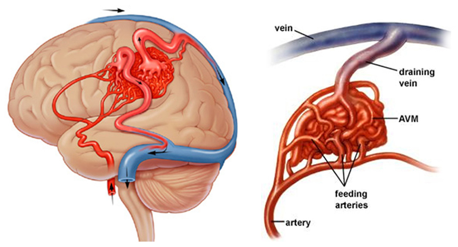
Fundamentally, a cerebral arteriovenous malformation refers to an abnormal tangle of vessels in the brain, where arteries are connected directly to veins. This creates a shunt where arterial blood bypasses the brain parenchyma and goes straight to the venous system. Typically this malformation will have feeding arteries, a nidus (nest of vessels comprised of both shunting arterioles and interconnected venous loops), and draining veins.
Here are some general features of this condition that might be appreciated across modalities:
- Tangled nest of vessels (nidus): this is the central location of the malformation. Its appearance can look like a “bag of worms” across imaging studies.
- Enlarged feeding vessels may be appreciated across varies studies (dilated arteries).
- Dilated draining vessels may be appreciated across varies studies. A venous varix may be present as well.
- Calcifications may be present in the nidus
COMPUTERIZED TOMOGRAPHY: NON-CONTRAST HEAD CT-SCAN
Key features of the appearance of cerebral cavernous malformation on this imaging modality are:
- Hyperdense appearance (relative to normal brain) on a non-contrast study (may be difficult to appreciate)
- Calcifications are best appreciated in the absence of contrast
- Signs of hemorrhage may be present and can be appreciated best on a non-contrast head CT.


COMPUTERIZED TOMOGRAPHY: IV CONTRAST HEAD CT-SCAN
Key features of the appearance of cerebral cavernous malformation on this imaging modality are:
- Contrast enhancing “bag of worms” appearance when IV contrast is used
- Enlarged feeding vessels/dilated draining vessels more clearly visualized due to the usage of contrast.


CT ANGIOGRAPHY (CTA)
Key features of the appearance of cerebral cavernous malformation on this imaging modality are:
- Contrast enhancing “bag of worms” appearance when IV contrast is used
- Enlarged feeding vessels/dilated draining vessels more clearly visualized due to the usage of contrast and also the ability to process the data and create vessel maps from the CTA.


MAGNETIC RESONANCE IMAGING: SUSCEPTIBILITY SEQUENCE HEAD MRI (GRE/SWI)
Key features of the appearance of cerebral cavernous malformation on this imaging modality are:
- No susceptibility artifact: there will not be an area of lost signal in the entire area of the malformation on GRE/SWI studies

MAGNETIC RESONANCE IMAGING: T1 WEIGHTED HEAD MRI
Key features of the appearance of cerebral cavernous malformation on this imaging modality are:
- Fast flow voids seen in the nidus: this can be appreciated on T1 imaging. Blood can move so quickly through the malformation that it does not return a signal to the MRI scanner (and will appear black).
- Incomplete contrast enhancement: some portions of the nidus may enhance partially, however much of the nidus will not enhance with contrast due to the speed of blood flow.


MAGNETIC RESONANCE IMAGING: T2 WEIGHTED HEAD MRI
Key features of the appearance of cerebral cavernous malformation on this imaging modality are:
- Fast flow voids seen in the nidus: this can be appreciated on various sequences, especially T2 weighted imaging. Blood can move so quickly through the malformation that it does not return a signal to the MRI scanner (and will appear black).

Page Updated: 08.09.2017