Page Contents
OVERVIEW
This page is dedicated to covering how the condition acute pancreatitis will appear on different types of radiological imaging studies.
BASIC CHARACTERISTICS
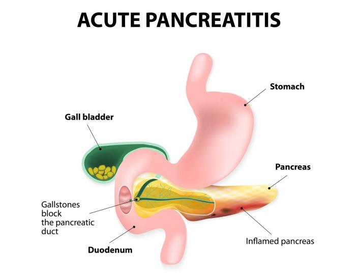
The central idea behind acute pancreatitis is that the pancreas (and very likely surround tissues) will be inflamed.

Here are some features of appendicitis that can be observed across all imaging studies:
- Enlargement of part of/all of the pancreas: the normal measurements for the pancreas are 3 cm for the head, 2.5 cm for the body, and 2 cm for the tail.
- Peripancreatic stranding: fat stranding can be present around the pancreas (which suggests inflammation).
- Peripancreatic fluid collections: given the disease process fluid may collect/be present around the pancreatic tissues.
- Pancreatic necrosis: low attenuation signals may be present in the pancreas due to necrosis. This is usually easier to visualize when IV contrast is used.
- Pseudocyst formation: this can be a complication of the acute pancreatitis. It is not present in every case. A fibrous tissue encapsulates a walled off collation of pancreatic juices. The wall can enhance with contrast.
COMPUTIRIZED TOMOGRAPHY (CT-SCAN)
MAGNETIC REASONANCE IMAGING (MRI)
Page Updated: 01.11.2017