Page Contents
OVERVIEW
This page is dedicated to covering how the condition appendicitis will appear on different types of radiological imaging studies.
BASIC CHARACTERISTICS
Appendicitis refers to acute inflammation of the appendix (outpouching of the large intestine).

Here are some features of appendicitis that can be observed across all imaging studies:
- Dilation: typically the key finding of an acute appendicitis is that it will be dilated/enlarged.
- Perforation: this can occur a certain percentage of the time. Periappendiceal extraluminal air or a periappendiceal abscess may be present.
ABDOMINAL ULTRASOUND
Here are some features of appendicitis that can be observed on abdominal ultrasound:
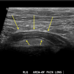
- Dilation: the appendix typically is dilated in acute appendicitis.
Click on the thumbnails below to open up a gallery of examples demonstrating appendicitis on ultrasound:
COMPUTIRIZED TOMOGRAPHY (CT-SCAN)
Here are some features of appendicitis that can be observed on CT scans:
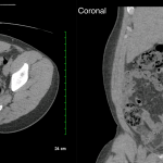
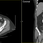
- Dilation of the appendix: typically > 6 mm is the size cutoff used to suspect appendicitis
- Periappendiceal inflammation: streaky, disorganized, linear, high signal densities in the surrounding fat tissues (can be referred to as fat stranding).
- Increased contrast enhancement in appendix wall (IV contrast): as a result of inflammation. This can be radiologically interpreted as wall thickening of the appendix.
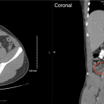
- Lumen of appendix not filled with contrast (Oral contrast): typically appendicitis is characterized by obstruction of the appendices lumen, so the lumen should not fill with oral contrast if it is used.
Click on the thumbnails below to open up a gallery of examples demonstrating appendicitis on CT scans without any contrast:
Click on the thumbnails below to open up a gallery of examples demonstrating appendicitis on CT scans with only IV contrast:
Click on the thumbnails below to open up a gallery of examples demonstrating appendicitis on CT scans with both IV and oral contrast:
MAGNETIC REASONANCE IMAGING (MRI)
Here are some features of appendicitis that can be observed on magnetic resonance imaging:
- Dilation: the appendix typically is dilated in acute appendicitis.
Page Updated: 01.10.2017
