OVERVIEW
This page is dedicated to covering the radiological appearance of an endotracheal tube.
WHAT IS IT?
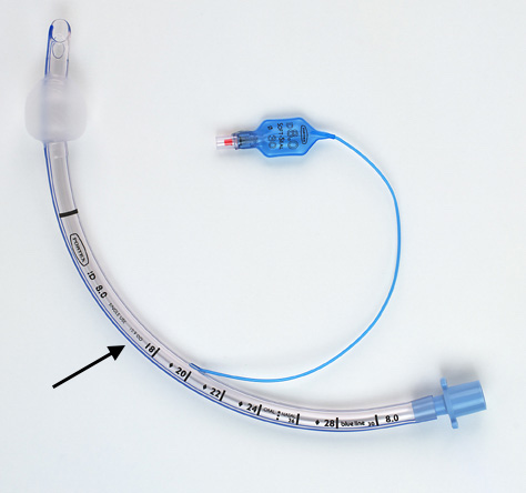
An endotracheal tube is a wide bored breathing tube that is used to assist ventilation. It typically is about 1cm in width, and has a radiopaque marker stripe on the side. The tip often is diagonally shaped. It is a piece of medical equipment that can be visualized on medical imaging.
WHAT TO LOOK FOR ON IMAGING
Often the proper placement of the endotracheal tube can be assessed on a chest X-ray:
Location of the tip: With the patient’s head in the neutral position (i.e. bottom of mandible is at the level of C5/C6) the tip of the tube should be about 3 to 5 cm from the carina. This is roughly half the distance between the medial ends of the clavicles and the carina. Neck flexion may cause a 2 cm decent of the tip which is why it should be a further distance from the carina.
Inflation of the cuff: the inflated cuff of the tube should NOT distend the lumen of the trachea.
ISSUES WITH TUBE PLACEMENT
Sometimes the improper placement of endotracheal tubes can be appreciated on a chest X-ray. Here is what to look out for:
Placement of the tip in the bronchus: most commonly these tips are malpositioned into he main or right lower lobe bronchi. This can lead to atelectasis of the non-aerated lung and pneumothorax can also be caused by damage to pulmonary structures.
Placement of the tip high in the neck: this can lead to damage of the vocal cords. The tube should be at least 3 cm distal to the vocal cords.
Intubation of the esophagus: if the tube was inserted in the esophagus the stomach will be hyper inflated.
Page Updated: 01.08.2016