Page Contents
OVERVIEW
This page is dedicated to covering the important radiological finding of a pneumomediastinum.
WHAT IS IT?
A pneumomediastinum refers to a finding on a chest X-ray where air is found in the mediastinum.
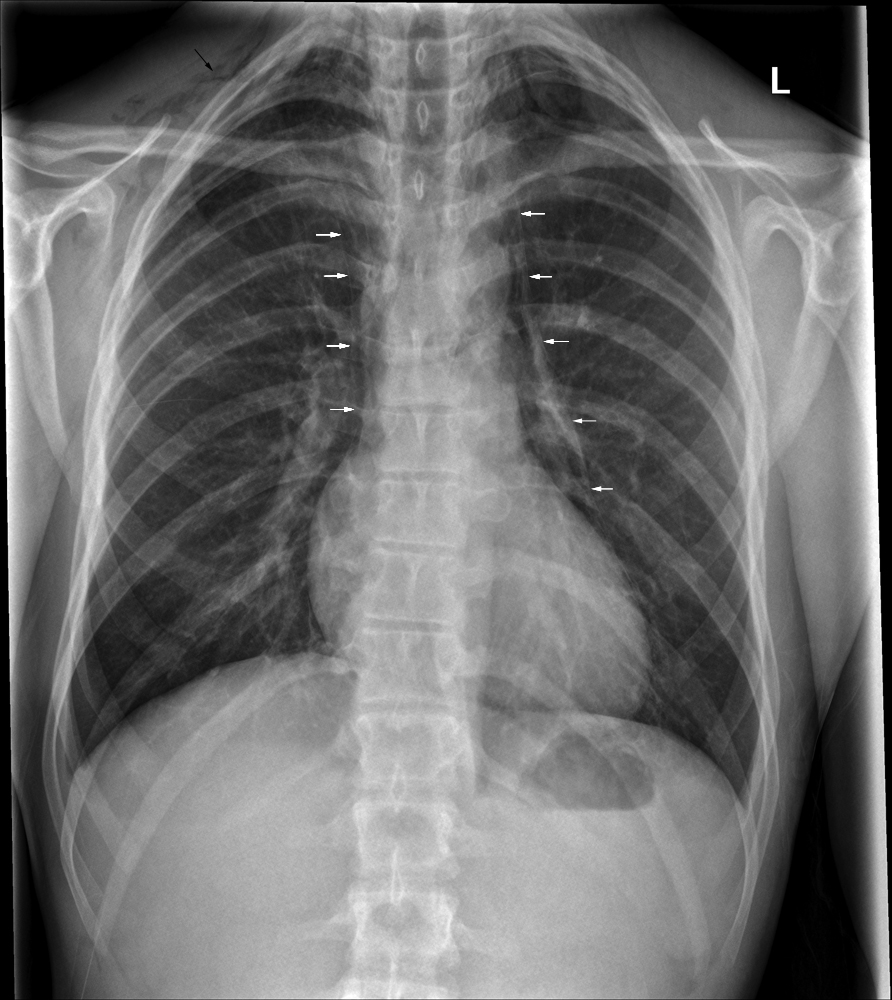
There can be a few different findings that are described as pneumomediastinum:
- Linear streak like lucency associated with a thin white line that is alongside the left heart border
- Streaky air outlining the great vessels (aorta, superior vena cava, carotid arteries)
- Linear streaks of air parallel to the spine in the upper thorax/neck (surround the esophagus and trachea)
DIFFERENTIAL DIAGNOSIS FOR THIS FINDING
When seeing a pneumomediastinum, it is important to keep in mind the following possible causes of this finding:
- Rupture of alveoli: can be caused by pulmonary interstitial emphysema
- Rupture of distal esophagus: this can be secondary to very forceful retching/vomiting (Boerhaave syndrome), instrumentation
- Rupture of the tracheobronchial tree: iatrogenic causes (traumatic intubation), penetrating wounds, severe blunt trauma.
- Pneumoperitoneum: secondary to things such as laparoscopy
KEY FEATURES TO LOOK FOR WHEN CHARACTERIZING THE FINDING
When seeing a pneumomediastinum, there are a few important radiological features one should look at to try and characterize the finding. These features can help navigate the differential diagnosis above.
Page Updated: 01.07.2016