OVERVIEW
This page is dedicated to covering how a subdural hemorrhage will appear on different types of radiological imaging studies.
BASIC CHARACTERISTICS
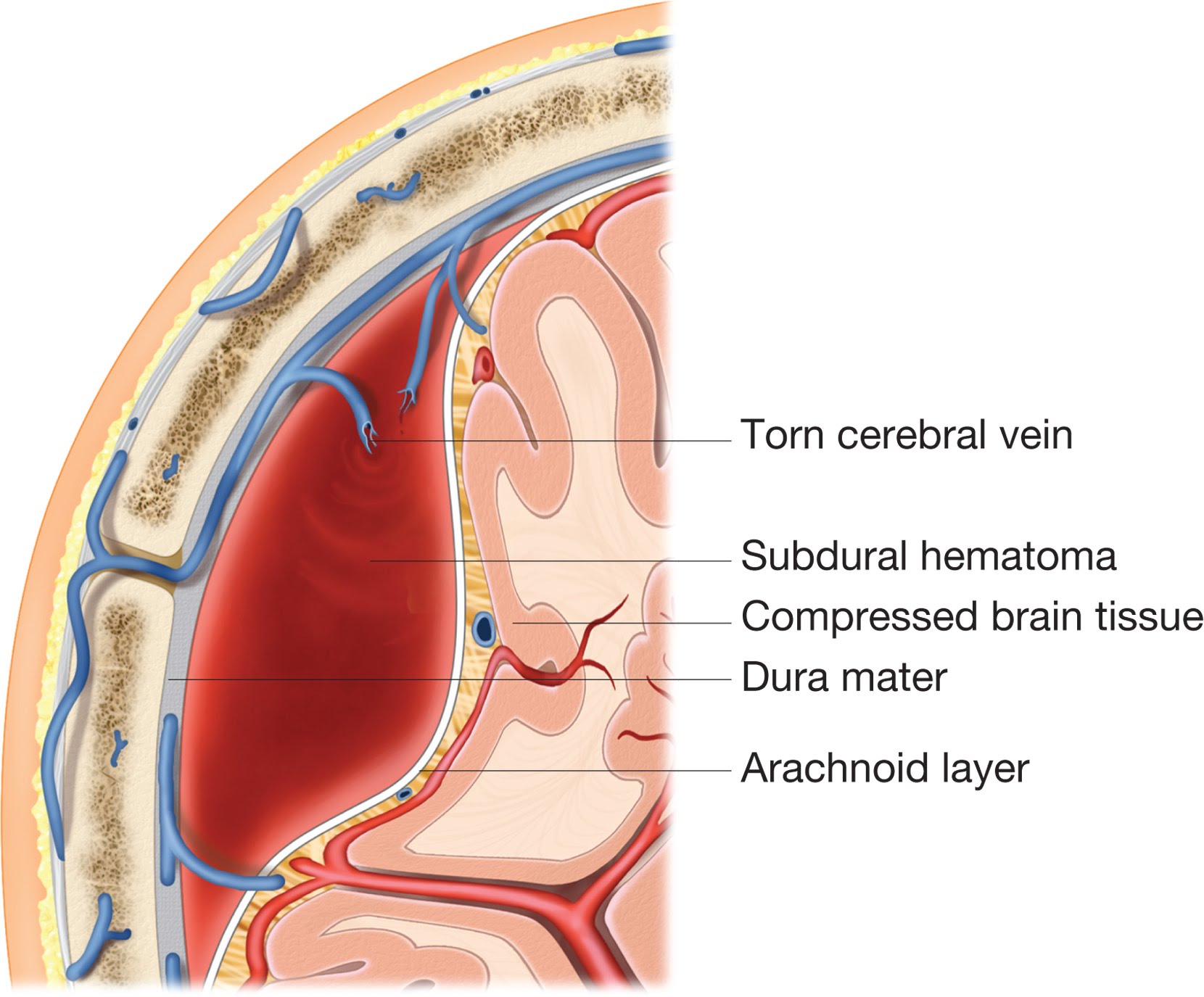
A subdural hematoma/hemorrhage refers to bleeding that occurs in the space underneath the dura but above arachnoid membranes. It can occur either in the brain or the spinal cord.
Here are some general radiological features of a subdural hemorrhage:
- Shape: the characteristic description of the shape of a subdural hemorrhage is “crescent-shaped”.
- Suture lines: the bleeding of a subdural hemorrhage will be able to cross the suture lines of the skull.
NON-CONTRAST HEAD CT SCAN
A non-contrast CT-scan is the preferred study used to evaluate for intracranial bleeding. Because of this, it is useful to appreciate how a subdural hemorrhage will appear on this scan. Make sure to review how to read non-contrast head CT scans, as well as the archive of unremarkable non-contrast head CT scans as a reference point.
The archive below organizes different examples of how a subdural hemorrhage will appear on a non-contrast head CT-scan. Click on the thumbnails below to view the archive.