OVERVIEW
This page is dedicated to covering how a epidural hematoma will appear on different types of radiological imaging studies.
BASIC CHARACTERISTICS
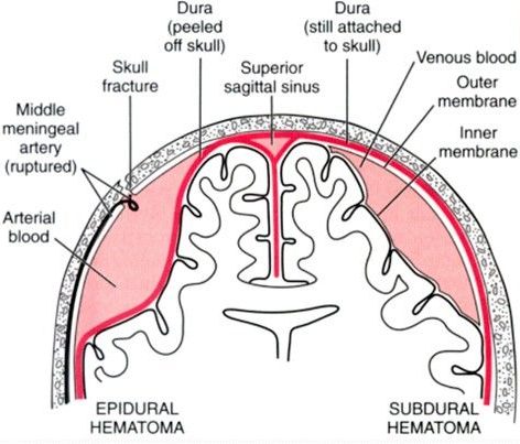
An epidural hematoma refers to bleeding that occurs between the dura meningeal layer and the skull (the bleeding will separate the dural from the bone. It is classically caused by the rupture of the middle meningeal artery, although more chronic presentations can be caused by venous bleeding. This is often secondary to fracture of the temporal bone.
Here are some general radiological features of a epidural hematoma:
- Shape: the characteristic description of the shape of a epidural hematoma is “lens-shaped”. Other similar descriptors that are used are biconvex or lentiform.
- Suture lines: the bleeding of a epidural hematoma will not cross the suture lines of the skull.
NON-CONTRAST HEAD CT SCAN
A non-contrast CT-scan is the preferred study used to evaluate for intracranial bleeding. Because of this, it is useful to appreciate how a epidural hematoma will appear on this scan. Make sure to review how to read non-contrast head CT scans, as well as the archive of unremarkable non-contrast head CT scans as a reference point.
The archive below organizes different examples of how a epidural hematoma will appear on a non-contrast head CT-scan. Click on the thumbnails below to view the archive.
Page updated: 10.11.2016