OVERVIEW
This page is dedicated to covering how the small intestine will appear across different radiological studies.
ABDOMINAL X-RAY
An abdominal X-ray is a very routinely ordered study, and a core component of reading this study is to evaluate the appearance of the small intestine.
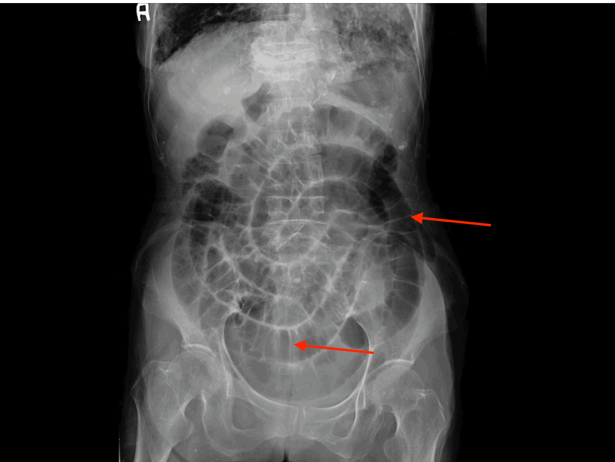
Characteristics of the small bowel on a KUB: the below features can help identify the small bowel on a KUB.
- Central location: generally these bowel loops will be located more in the central region of the abdomen (compared to the large intestine)
- Presence of air: there are generally 2-3 locations that are air filled in a “normal” small bowel.
- Air/fluid levels: three or less fluid levels are within the range of normal
- Diameter: the normal diameter of the small bower is < 2.5 cm (~ 1 inch). When maximally dilated it is about 5.0 cm.
- Valvulae: these are folds of the small bowel that span the entire lumen, and can appear clearly as “white lines” on the film (intestinal markings).
Page Updated: 10.07.2016