Page Contents
OVERVIEW
This page is dedicated to covering how the condition small bowel obstruction will appear on different types of radiological imaging studies. Feel free to review the page that covers the radiological anatomy of the small intestine.
BASIC CHARACTERISTICS OF A SMALL BOWEL OBSTRUCTION
Fundamentally a small bowel obstruction is a very self descriptive condition, and is caused by some process that impedes the flow of material through the small bowel. The specific cause can be variable (truly any obstructive process can cause it) however by the nature of this condition, it is important to notice that there are some unifying characteristics that can be observed throughout radiological studies.
Here are some features of a small bowel obstruction that can be observed across all imaging studies:
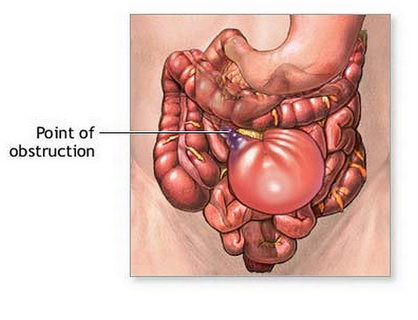
Location: this condition will occur at some point along the lumen of the small intestine.
Proximal to the obstruction bowel loops will be dilated
Distal to the obstruction bowel loops will be decompressed
SUPINE ABDOMINAL X-RAY (KUB)
An abdominal X-ray can be one of the first studies ordered when a SBO is suspected. The supine view will be one of the images collected.
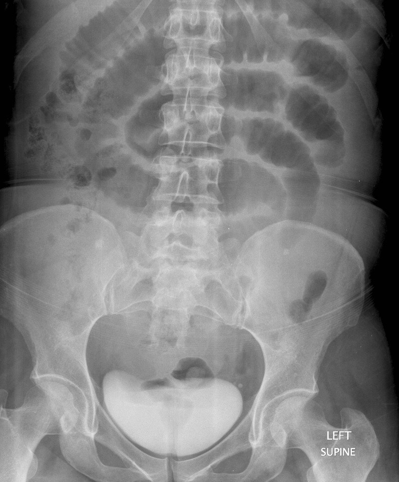
Some key features to keep in mind for the appearance of a SBO on a supine KUB
- Dilated bowel loops: the normal intestine is < 2.5 cm in diameter. This diameter will be increased in patients with a SBO.
- Loss of valvulae: these are folds of the small bowel that span the entire lumen, and can appear clearly as “white lines” on the film (intestinal markings). They can be lost as the bowels dilate.
- Air/fluid levels: given that this patient’s orientation is supine, these air/fluid levels generally are not seen on this view.
- Gas in sigmoid/rectum: often gas may be absent in the sigmoid colon or rectum, because it can not move past the obstruction.
UPRIGHT ABDOMINAL X-RAY (KUB)
An abdominal X-ray can be one of the first studies ordered when a SBO is suspected. The upright view will be one of the images collected.
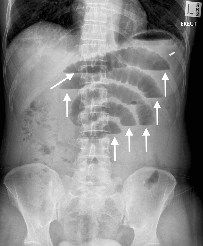
Some key features to keep in mind for the appearance of a SBO on a upright KUB
- Dilated bowel loops: the normal intestine is < 2.5 cm in diameter. This diameter will be increased in patients with a SBO.
- Loss of valvulae: these are folds of the small bowel that span the entire lumen, and can appear clearly as “white lines” on the film (intestinal markings). They can be lost as the bowels dilate.
- Air/fluid levels: these can be most easily seen on the upright film. More then 3 air/fluid levels is concerning for a process like and SBO.
- Gas in sigmoid/rectum: often gas may be absent in the sigmoid colon or rectum, because it can not move past the obstruction.
COMPUTIRIZED TOMOGRAPHY (CT-SCAN)
An abdominal CT scan will ultimately be the means by which a small bowel obstruction is more definitively diagnosed.
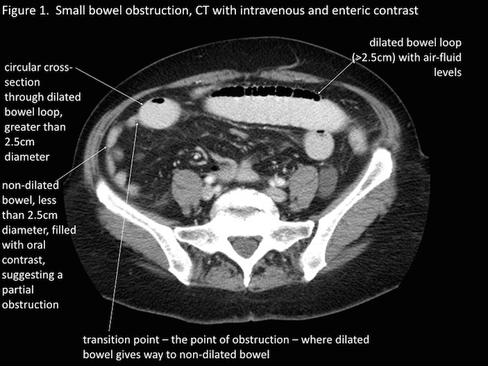
Key features characteristic of detecting an SBO on a CT-scan are:
- Proximal to the obstruction: dilated bowel loops proximal to the obstruction
- Distal to the obstruction: decompressed bowel loops distal to the obstruction
- The point of obstruction will be marked by this transition of dilated to decompressed bowel.
- Air/fluid levels: these can be seen proximal to the obstruction (easiest to appreciate on the axial view).
Page Updated: 09.21.2016