Page Contents
OVERVIEW
This page is dedicated to covering the differential diagnosis for chest pain. What’s more, the possible diagnosis that present with chest pain will be organized by diagnostic modality.

WHAT ARE THE MAJOR CATEGORIES OF DIAGNOSTIC MODALITIES THAT ARE USED?
When thinking about the differential for chest pain the following diagnostic modalities come to mind:
- History and Physical
- Venous blood studies
- Electrocardiogram
- Chest X-ray
- Endoscopy
DIAGNOSIS MADE WITH THE HISTORY AND PHYSICAL
Some causes of chest pain might not really require much a of a clinical workup (and are really more of a “clinical diagnosis” largely made from the history). Examples of these types of conditions that cause chest pain are listed below.
- Gastroesophageal Reflux Disease (GERD): really the history of present illness will help diagnose this condition (although one generally will have to work up other possible causes as this may need to be a diagnosis of exclusion).
- Muscle strain: the history and physical will be very useful in deciding if the pain in the chest is from an isolated muscle strain (for example, physically poking the chest will increase the pain felt).
- Herpes zoster: the reactivation of this virus in dermatomes across the chest can manifest and chest pain. This diagnosis is made by physically examining the chest.
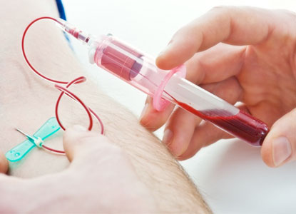
VENOUS BLOOD STUDIES
Venous blood studies are a very foundational component of a clinical workup. This is no different in the case of chest pain, and it is important to appreciate what information these studies can tell us (and what diseases can be implicated/diagnosed by them).
Cardiac biomarkers (i.e. troponin): these types of studies can help diagnose the following.
- Myocardial infarction (STEMI, NSTEMI)
Serum lipase (and amylase): the elevation of these enzymes in the serum can suggest
ELECTROCARDIOGRAM
In the realm of chest pain, the threshold for order an EKG is very low! It may have even been performed before the patient comes into the clinic.
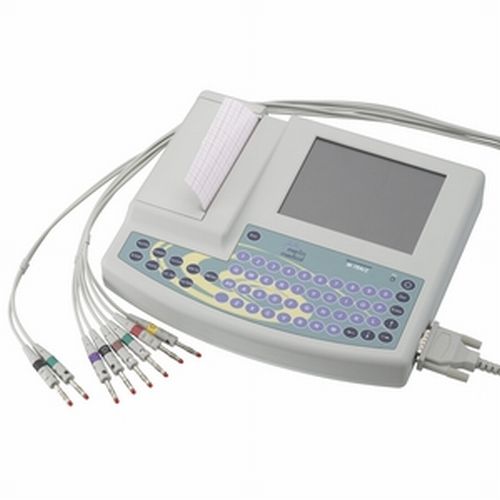
Given how commonly an EKG is ordered for the workup of chest pain, it is important to appreciate what disease processes can be implicated by results from this test.
EKG at rest:
- Myocardial infarction (STEMI, NSTEMI) of course can be implicated/diagnosed with this study (ST elevations, ST depressions, T wave inversions depending on the context).
- Pericarditis can be implicated (widespread concave ST elevations and PR depressions throughout most of the leads)
- Unstable angina may be detected with the presence of ST depressions or T-wave inversions
Stress EKG:
- Stable angina can be detected when the patient is exerting themselves.
CHEST X-RAY
Very likely this study might be ordered (especially in the context of certain physical exam findings).
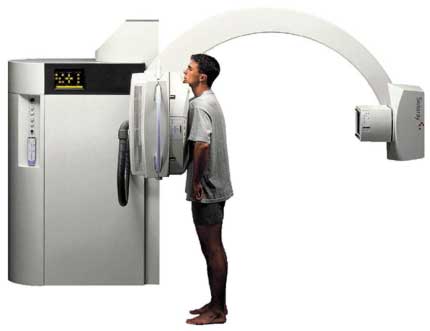
When thinking about chest pain, a chest X-ray can help implicate the following disease processes by revealing the following findings:
- Widened mediastinum: this can suggest disease processes such as an aortic dissection, thoracic aortic aneurysm,
- Pericardial effusion can be implicated on a chesty X-ray (by the presence of extra fluid around the heart/classic “water bottle” shape of heart).
- Pneumothorax can be diagnosed by witnessing deceased lung markings on the film. Signs of a tension pneumothorax can also be present in certain cases.
- Pneumonia can be diagnosed on an X-ray (especially in the right clinical setting). With this in mind, it can be difficult to label a pneumonia the cause of chest pain before ruling out other more likely causes.
- Pleural effusion may also be the cause of the patients chest pain, and is often diagnosed on this study. Look here for more examples of how a pleural effusion will look on a chest X-ray.
- Esophageal perforation might be suggested by the findings of: mediastinal air, pleural effusion, pneumothorax, and subdiaphragmatic air
- Rib fracture can cause chest pain, and can be visualized on a plain film study
ENDOSCOPY
Sometimes it is important to visualize directly what is happening along the GI tract.
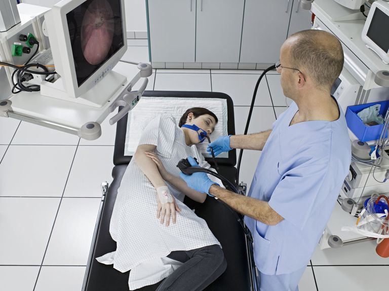
While there are not many causes of chest pain diagnosed by upper endoscopy, there are a few worth noting below:
- Peptic ulcer disease: visualizing the suspected ulcers really is the gold standard for diagnosing this condition.
Page Updated: 07.22.2016