Page Contents
- 1 WHAT IS IT?
- 2 WHAT CAUSES IT?
- 3 WHY IS IT A PROBLEM?
- 4 WHAT MAKES US SUSPECT IT?
- 5 HOW DO WE CONFIRM A DIAGNOSIS?
- 6 HOW DO WE RULE OTHER DIAGNOSES OUT?
- 7 HOW DO WE TREAT IT?
- 8 HOW WELL DO THE PATIENTS DO?
- 9 WAS THERE A WAY TO PREVENT IT?
- 10 WHAT ELSE ARE WE WORRIED ABOUT?
- 11 OTHER HY FACTS?
- 12 ARCHIVE OF STANDARDIZED EXAM QUESTIONS
- 13 FURTHER READING
- 14 Page Updated: 07.12.2016
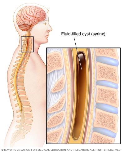
WHAT IS IT?
A syringomyelia is a cystic cavity (syrinx) within the spinal cord (referred to as cystic degeneration of the spinal cord). It is referred to as a hydromyelia if found in the central canal.
WHAT CAUSES IT?
This condition can be congenital, is associated with Arnold-Chiari malformation, and can also arise after trauma, a tumor, or meningitis.
WHY IS IT A PROBLEM?
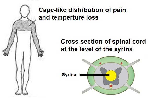
As this cavity enlarges it can damage structures in the spinal cord. Often the anterior spinal commissural fibers are disrupted (leading to the patterns of sensory loss described below). As the cavity enlarges other neurological findings can be detected.
WHAT MAKES US SUSPECT IT?
Risk factors: Arnold-Chiari malformations, trauma, tumors
Sensory loss (pinprick and temperature) that is often bilateral and “cape-like’ in its distribution. Fine touch sensation is typically preserved because it does not cross at the anterior commissure.
Muscle atrophy/weakness/pain can occur as the anterior horn becomes affected. This will also present with decreased muscle tone and decreased reflexes.
Horner syndrome can be caused by late stage syringomyelia. This consists of the characteristic triad of unilateral:
- Miosis
- Ptosis
- Anhydrosis
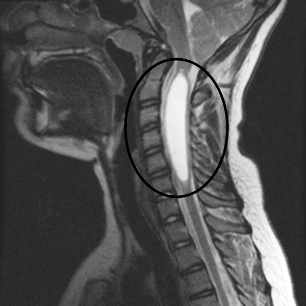
HOW DO WE CONFIRM A DIAGNOSIS?
MRI can visualize the syringomyelia cavity in the spinal cord.
HOW DO WE RULE OTHER DIAGNOSES OUT?
The sensory loss patterns will help rule out ALS (which typically only has motor signs).
HOW DO WE TREAT IT?
Surgical correction
HOW WELL DO THE PATIENTS DO?
Often patients will not improve after surgical intervention.
WAS THERE A WAY TO PREVENT IT?
N/A
WHAT ELSE ARE WE WORRIED ABOUT?
Arnold Chiari Malformation: this is associated with syringomyelia and meningomyelocele. It is congenital extension of the cerebellar tonsils through the foramen magnum. It can obstruct the flow of CSF and result in hydrocephalus.
Malignancy can sometimes be the underlying cause of this condition.
OTHER HY FACTS?
Most commonly occurs at C8-T1 level in spinal cord.
ARCHIVE OF STANDARDIZED EXAM QUESTIONS
This archive compiles standardized exam questions that relate to this topic.