This page is dedicated to providing an organized archive of images that demonstrate the appearance of a cranial meningioma. This archive is organized by the specific imaging study utilized to image the cranial meningioma.
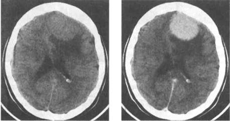
CT scan of a cranial meningioma before (left pane) and after (right pane) the administration of IV contrast (source)
T1-WEIGHTED MRI
Example # 1:
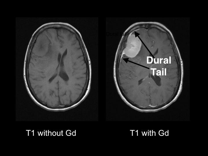
Appearance of meningioma on a T1 weighted MRI image before and after contrast enhancement. The characteristic “dural tail” can be seen on the right pane with enhancement (source).
Example #2:
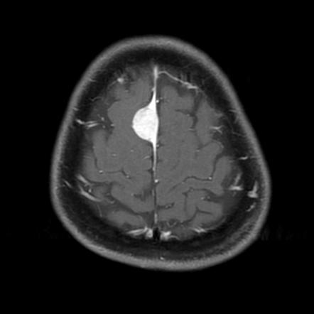
Axial T1 weighted MRI showing a parasagittal meningioma with a characteristic dural tail (source)
Example #3:
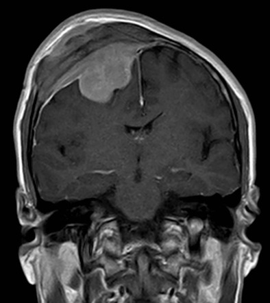
T1 weighted MRI with contrast showing meningioma with characteristic dural tail (source)
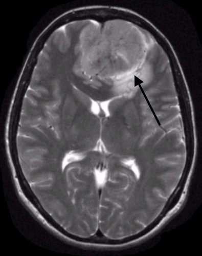
T2-WEIGHTED MRI
Example #1:
T2 axial image of a cranial meningioma. The arrow points to the presence of CSF in the “cleft” between the tumor and brain parenchyma. This is a feature that is characteristic of a meningioma (source)