Page Contents
OVERVIEW
This page is dedicated to covering how a cranial meningioma will appear on different types of radiological imaging studies.
BASIC CHARACTERISTICS OF A MENINGIOMA
We have to appreciate that fundamentally a meningioma is a brain tumor that arises from the meningeal layers (most specifically from the arachnoid cap cells of leptomeninges) covering the brain and NOT the actual brain parenchyma itself. This etiology of this mass will dictate its general appearance on imaging.
Here are some features of a meningioma that can be observed across all imaging studies:
Location: typically most of these tumors are supratentorial in anatomical location.
Origin of tumor: given the etiology of a meningioma, the tissue will arise from the meninges (“dural based) that surround the brain, and the mass will be extra-axial (outside, brain parenchyma). A dural attachment (sometimes called a “dural tail”) may be appreciated depending on the study used.
Homogeneous nature: most meningiomas will generally enhance homogeneously/uniformly on imaging studies
Trapped CSF: given the nature of the tumor, CSF will often be “trapped” in the area (or “cleft”) between the tumor and the brain.
COMPUTIRIZED TOMOGRAPHY (CT-SCAN)
A CT scan may be the first time a cranial meningioma is detected:
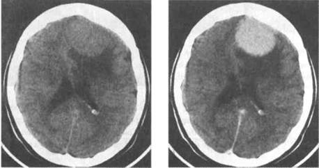
Some key features to keep in mind for the appearance of meningioma on CT scan are:
- Mass characteristics: typically a sharply defined mass that originates from the meninges.
- Signal intensity: most are hyper-intense, the rest generally are iso-intense
- Contrast enhancement: with the addition of IV contrast most meningiomas will enhance homogeneously
MAGNETIC REASONANCE IMAGING (MRI)
There are of course many different types of MRI sequences each with their own characteristics that are consistent with the presentation of a cranial meningioma.
T1 weighted image: many of the basic features of a cranial meningioma will be present on a T1 MRI sequence.
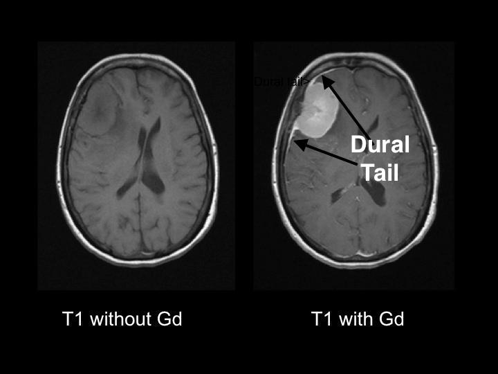
Notable T1 characteristics:
- Intensity: typically iso-/hypo-intense in nature (relative to the cortex of the brain). This is BEFORE contrast is added.
- Enhancement: this tumor type will normally enhance homogeneously with the addition of galladinium.
- Dural tail: this feature of the meningioma can be seen after contrast enhancement (such as in the image above). This feature actually is best visualized on a FLAIR MRI sequence.
T2 weighted image: many of the basic features of a cranial meningioma will be present on a T2 MRI sequence.
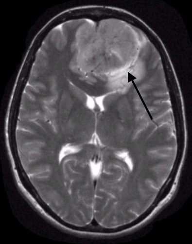
Notable T2 characteristics:
- CSF in the “cleft”: given the nature of a T2 weighted image, this is the best MRI series to visualize the presence of CSF in the cleft between the tumor tissue and the brain parenchyma. This is a feature characteristic of cranial meningiomas.
FURTHER READING
Archive Of Radiology Images: Cranial Meningioma Organized By Imaging Study
Archive Of Radiology Images: Cranial Meningioma Findings
Page Updated: 09.28.2016