Page Contents
OVERVIEW
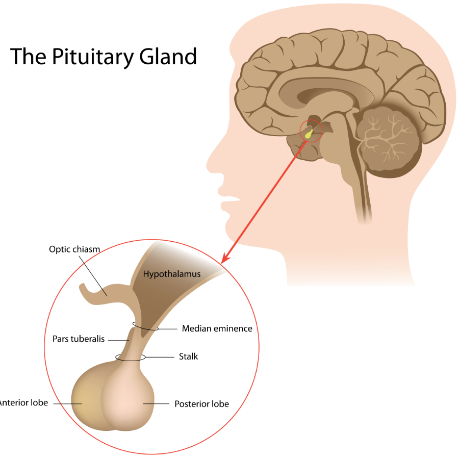
This page is dedicated to covering how the pituitary gland will appear across different radiological studies. The pituitary gland is an important endocrinological organ that is found at the base of the skull.
CLINICAL SIGNIFICANCE
Pituitary gland is clinically significant because it is responsible for storing/producing many different important hormones. It can also be the site of the formation of malignancy.
APPEARANCE OF THIS STRUCTURE ON T1-WEIGHTED MRI IMAGING (SAGITTAL CROSS SECTIONS)
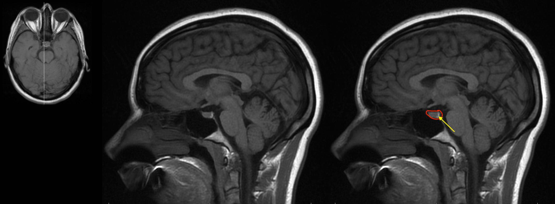
The pituitary gland can be appreciated on sagittal MRI cross sections by both its location (it sits in the sella turcica/pituitary fossa at the base of its skull) as well as by the presence of a characteristic T1 intense “bright spot” found in the posterior pituitary gland. This bright spot is thought to be caused by the presence of vasopressin in the posterior pituitary. The image below helps show a characteristic example of this.
HERE IS A GALLERY OF MORE EXAMPLES. CLICK ON THE THUMBNAILS BELOW TO OPEN UP THE GALLERY.


APPEARANCE OF THIS STRUCTURE ON T1-WEIGHTED MRI IMAGING (AXIAL CROSS SECTIONS)
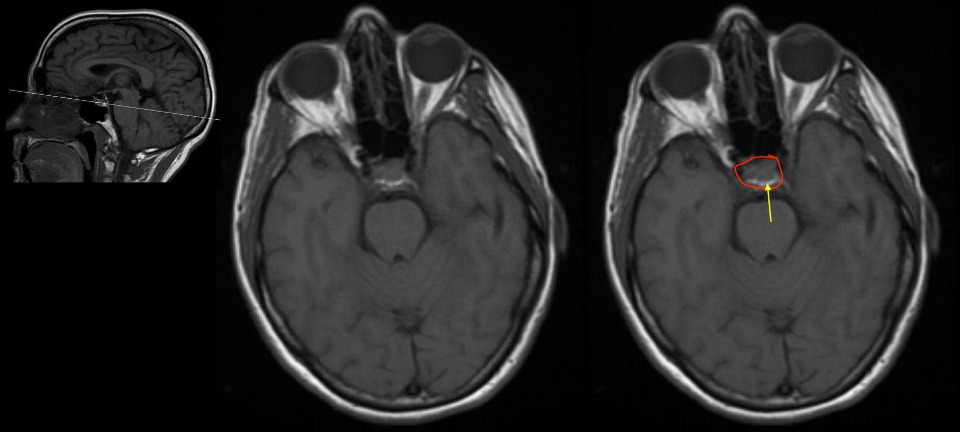
The pituitary gland can be appreciated on axial MRI cross sections as well. On T1 weighted imaging the characteristic T1 intense “bright spot” can be visualized. The image below helps demonstrate this concept.
HERE IS A GALLERY OF MORE EXAMPLES. CLICK ON THE THUMBNAILS BELOW TO OPEN UP THE GALLERY.


APPEARANCE OF THIS STRUCTURE ON T1-WEIGHTED MRI IMAGING (PRE AND POST CONTRAST)
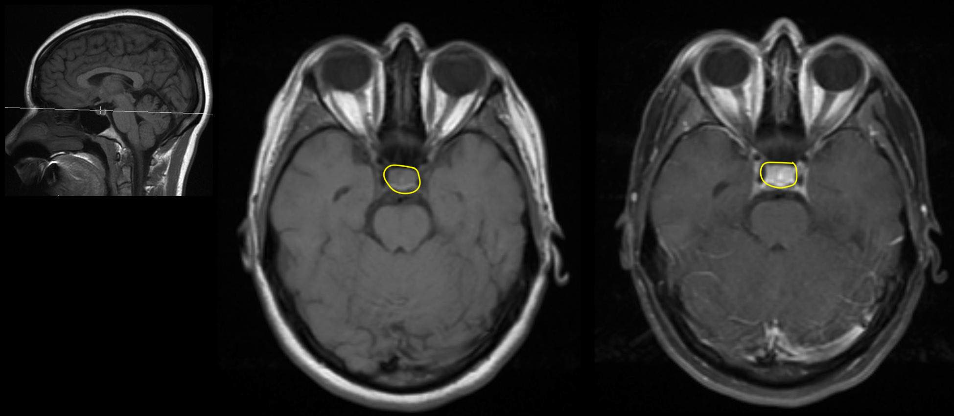
The pituitary gland can is a structure that will enhance on MRI imaging with the administration of contrast. The examples below will help demonstrate this.
HERE IS A GALLERY OF MORE EXAMPLES. CLICK ON THE THUMBNAILS BELOW TO OPEN UP THE GALLERY.


ACKNOWLEDGEMENTS
A very special thanks goes to Dr. Pierre Sasson who made this page possible with his expertise and insight.
Page Updated: 01.23.2018