OVERVIEW
This page is dedicated to covering how an subarachnoid hemorrhage will appear on different types of radiological imaging studies.
BASIC CHARACTERISTICS
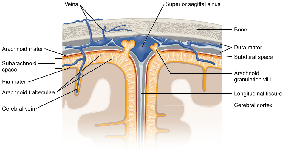
A subarachnoid hemorrhage (SAH) refers to bleeding that occurs in the subarachnoid space between the arachnoid membrane and the pia matter that surrounds the brain. It can be caused by the rupture of an intracranial aneurysm (such as a saccular or berry aneurysm).
NON-CONTRAST HEAD CT SCAN
A non-contrast CT-scan is the preferred study used to evaluate for intracranial bleeding. Because of this, it is useful to appreciate how a subarachnoid hemorrhage will appear on this scan. Make sure to review how to read non-contrast head CT scans, as well as the archive of unremarkable non-contrast head CT scans as a reference point.
The archive below organizes different examples of how a subarachnoid hemorrhage will appear on a non-contrast head CT-scan. Click on the thumbnails below to view the archive.