Page Contents
- 1 WHAT IS IT?
- 2 WHAT CAUSES IT?
- 3 WHY IS IT A PROBLEM?
- 4 WHAT MAKES US SUSPECT IT?
- 5 HOW DO WE CONFIRM A DIAGNOSIS?
- 6 HOW DO WE RULE OTHER DIAGNOSES OUT?
- 7 HOW DO WE TREAT IT?
- 8 HOW WELL DO THE PATIENTS DO?
- 9 WAS THERE A WAY TO PREVENT IT?
- 10 WHAT ELSE ARE WE WORRIED ABOUT?
- 11 OTHER HY FACTS?
- 12 ARCHIVE OF STANDARDIZED EXAM QUESTIONS
- 13 FURTHER READING
WHAT IS IT?
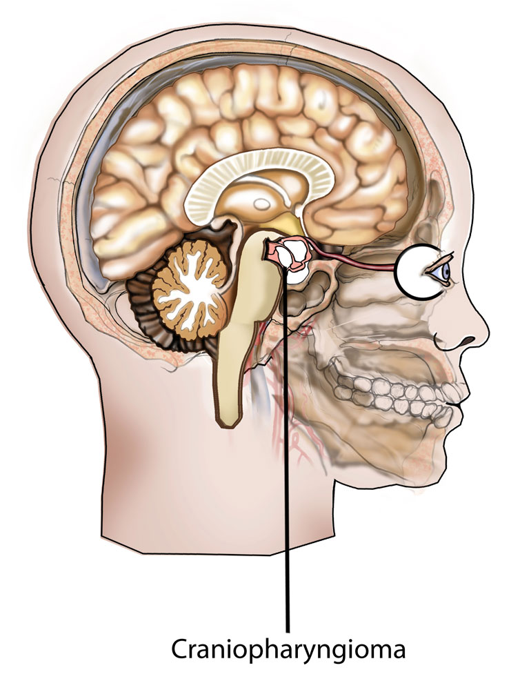
A craniopharyngioma is a benign suprasellar brain tumor that often occurs in children. It is a very rare tumor.
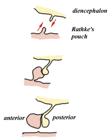
WHAT CAUSES IT?
This tumor arises from the epithelial remnants of Rathke’s pouch. Rathke’s pouch is a developmental structure during embryogenesis. It is a depression in the roof of the development mouth, and gives rise to the anterior pituitary.
WHY IS IT A PROBLEM?
This tumor can compress on surrounding structures leading to damage/dysfunction:
- Visual field defects can be caused by compression of the optic nerve/optic chiasm.
- Hypopituitarism (decreased secretion of pituitary hormones) due to the mass effect or pituitary apoplexy.
WHAT MAKES US SUSPECT IT?
Risk factors
No clear risk factors. Most commonly diagnosed in children ages 5-15.
Initial Presentation
Common Chief Complaints:
- Headache can be common
- Vision loss can be caused by compression of optic nerve/optic chiasm.
- Growth retardation in children.
Associated Symptoms:
- Increased thirst/urination due to loss of ADH signaling
- Hearing loss
- Nausea/vomiting
Physical Exam Findings
Patient height may be decreased (growth charts in the low percentages) due to hypopituitarism.
Visual field examination can reveal bitemporal hemianopia (due to compression of the optic chiasm)
HOW DO WE CONFIRM A DIAGNOSIS?
Imaging (both CT and MRI) can show the tumor. Calcifications are commonly seen in this tumor type as they are commonly filled with cholesterol.
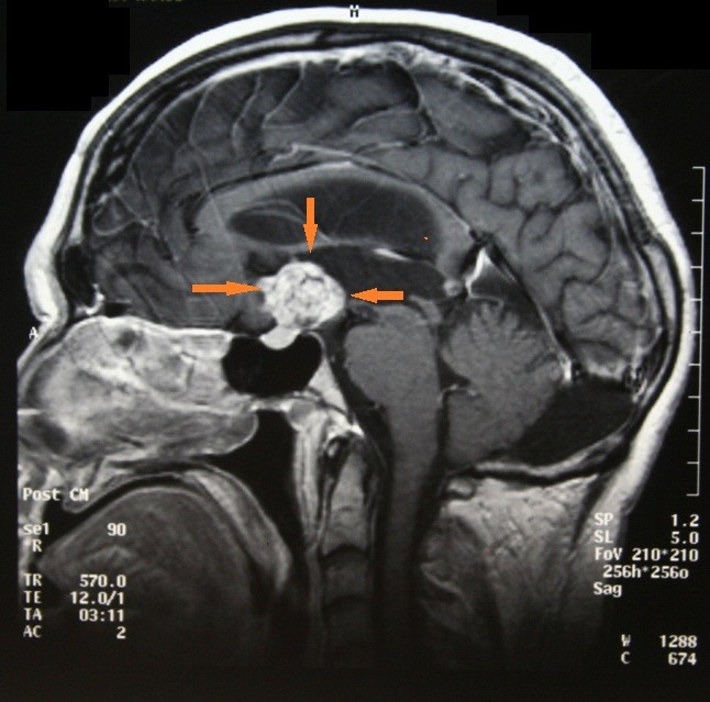
HOW DO WE RULE OTHER DIAGNOSES OUT?
MRI imagining: This tumor is often confused with pituitary adenoma. One distinguishing element (especially on standardized exams) is that calcifications are present in the craniopharyngioma (but are not really characteristic of the pituitary adenoma).
HOW DO WE TREAT IT?
Surgical resection is a common route for treatment. There are different surgical options depending on the nature of the tumor.
HOW WELL DO THE PATIENTS DO?
This tumor is generally benign, but often recurs after resection.
WAS THERE A WAY TO PREVENT IT?
N/A
WHAT ELSE ARE WE WORRIED ABOUT?
Permanent damage to cranial structures can occur if the tumor is allowed to grow and compress surrounding structures.
OTHER HY FACTS?
The ventromedial area of the hypothamlas can be destroyed by a craniopharyngioma. This area controls satiety, so its destruction can result in excessive eating.
Craniopharyngioma is the most common childhood supratentorial tumor.
ARCHIVE OF STANDARDIZED EXAM QUESTIONS
This archive compiles standardized exam questions that relate to this topic.
FURTHER READING
Page Updated: 07.20.2016