OVERVIEW
This page is dedicated to discussing how to find the appendix on imaging studies so that it can be evaluated. This is especially useful when trying to evaluate whether or not a patient may have acute appendicitis.
IMPORTANT LANDMARKS FOR LOCALIZING THE APPENDIX
The appendix is a small structure, that sometimes can be elusive. It is for this reason that having larger landmarks to try and help localize is useful!
It is important to realize that anatomy varies, and appendix location can also be variable. But with this in mind the following landmarks help narrow the search field for the appendix:
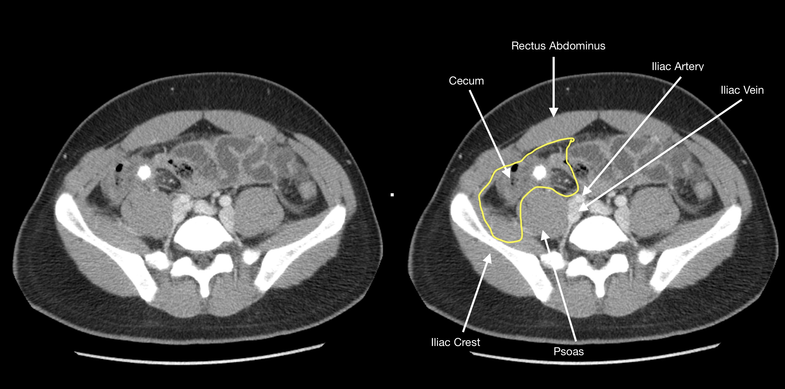
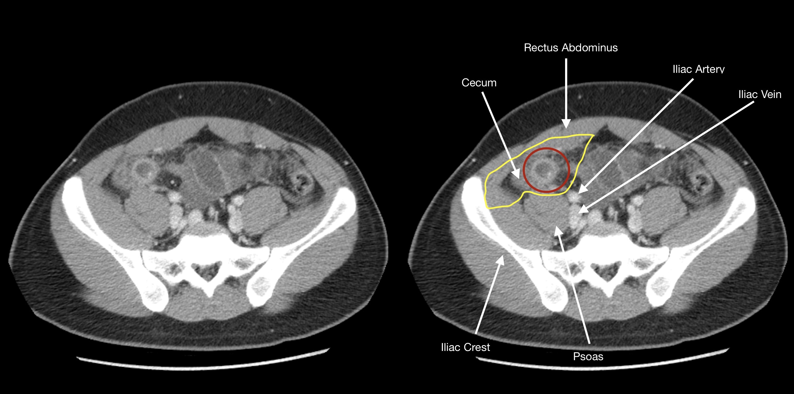
- The iliac crest: this is an easy to identify structure. Depending on the level it will represent either the posterior or both the posterior and lateral borders of where you must search to identify the appendix.
- The rectus abdominis; this muscle is also relatively easy to locate. It will represent the anterior border of the search field for the appendix.
- The psoas major: another clear muscular landmark, it will often be right next to the appendix. It commonly represents the posterior/medial border of the search field for the appendix.
- The common iliac artery/vein: these vessels will often represent the medial boundary for the search field for the appendix. It is important to appreciate however that depending on the level of the appendix in the superior/inferior plane this border may be the vessels proximal or distal to the common iliac artery/vein.
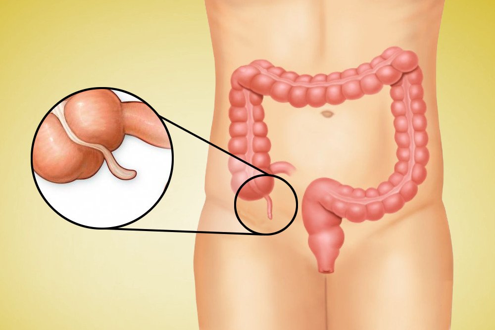
- The cecum: perhaps the most important landmark, the cecum will be adjacent to the appendix because the appendix originates from the cecum! Their relationship to one another can be variable however given that the appendix can be retrocecal etc.
LOCALIZING THE APPENDIX ON CROSS-SECTIONAL IMAGING (AXIAL PLANE)
Perhaps the most intuitive way to utilize the landmarks described above is to show how they are appleid to help find an appendix in a patient with acute appendicitis. A good initial place to start is to view cross sectional imaging in the axial plane.
Page Updated: 08.05.2019