Page Contents
OVERVIEW
This page is dedicated to covering how the calcarine sulcus will appear across different radiological studies. It is an important anatomical structure in the brain.
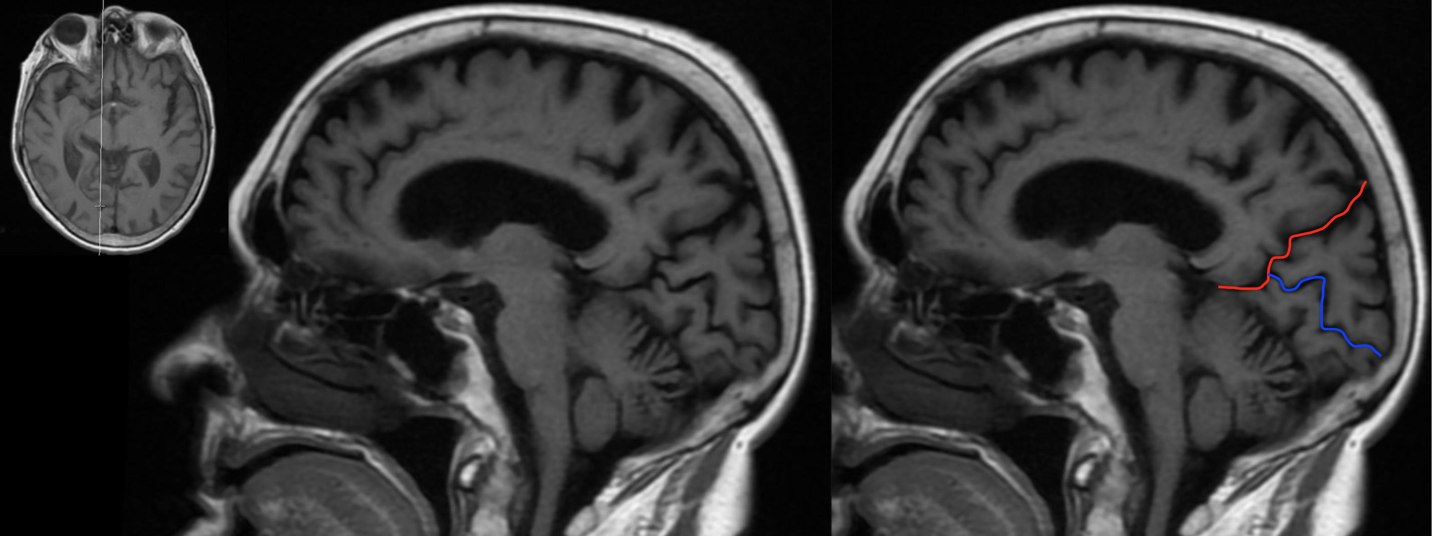
FINDING THE CALCARINE SULCUS ON SAGITTAl CROSS SECTIONS: Y-SIGN
Often times on sagittal sections near the midline of the brain, the parieto-occipital sulcus will join with the calcarine sulcus to form what looks like a “Y”. The image below depicts shows this characteristic “Y-sign”.
HERE ARE SOME EXAMPLES OF THIS SIGN SEEN ON A NON-CONTRAST HEAD CT-SCAN. CLICK THE THUMBNAILS TO VIEW THEM.


HERE ARE SOME EXAMPLES OF THIS SIGN SEEN ON A T1 WEIGHTED HEAD MRI WITHOUT CONTRAST. CLICK THE THUMBNAILS TO VIEW THEM.


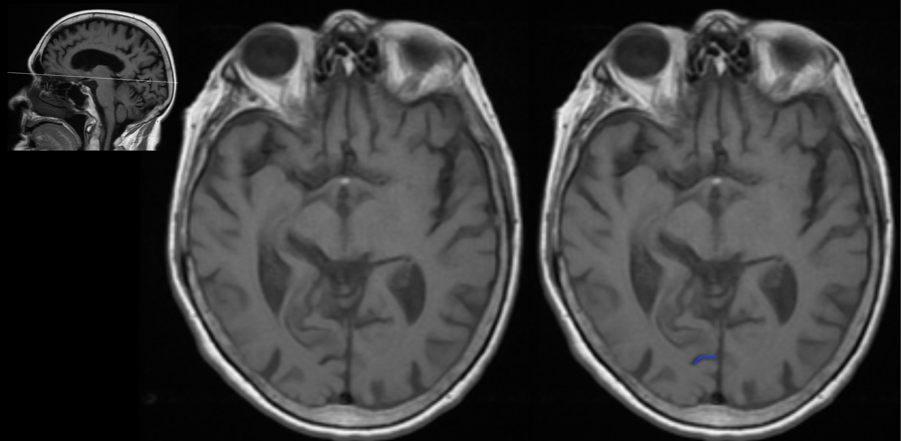
APPEARANCE OF THE CALCARINE SULCUS ON AXIAL CROSS SECTIONS
The calcarine sulcus can be seen on axial sections of the brain, however there is not a clear characteristic appearance of this structure on this section. Looking at the sagittal cross sections of the brain will help to locate this sulcus on axial images. An example of how this sulcus looks on axial imaging is shown below.
HERE ARE SOME EXAMPLES OF THIS SULCUS SEEN ON A NON-CONTRAST HEAD CT-SCAN (AXIAL ORIENTATION). CLICK THE THUMBNAILS TO VIEW THEM.


HERE ARE SOME EXAMPLES OF THIS SULCUS SEEN ON A T1 WEIGHTED HEAD MRI WITHOUT CONTRAST (AXIAL ORIENTATION). CLICK THE THUMBNAILS TO VIEW THEM.


ACKNOWLEDGEMENTS
A very special thanks goes to Dr. Pierre Sasson who made this page possible with his expertise and insight.
Page Updated: 11.25.2017