Page Contents
- 1 OVERVIEW
- 2 FINDING THE CENTRAL SULCUS ON AXIAL CROSS SECTIONS: OVERVIEW OF SIGNS
- 3 FINDING THE CENTRAL SULCUS ON AXIAL CROSS SECTIONS: SUPERIOR FRONTAL SULCUS SIGN (PRE-CENTRAL SULCUS SIGN)
- 4 FINDING THE CENTRAL SULCUS ON AXIAL CROSS SECTIONS: SIGMOID HOOK SIGN
- 5 FINDING THE CENTRAL SULCUS ON AXIAL CROSS SECTIONS: PARS BRACKET SIGN
- 6 FINDING THE CENTRAL SULCUS ON AXIAL CROSS SECTIONS: BIFID POST-CENTRAL SULCUS SIGN
- 7 FINDING THE CENTRAL SULCUS ON AXIAL CROSS SECTIONS: THIN POST-CENTRAL GYRUS SIGN
- 8 FINDING THE CENTRAL SULCUS ON AXIAL CROSS SECTIONS: INTRAPARIETAL SULCUS SIGN
- 9 FINDING THE CENTRAL SULCUS ON SAGITTAL CROSS SECTIONS: CINGULATE SULCUS/PARS MARGINALIS SIGN
- 10 ACKNOWLEDGEMENTS
OVERVIEW
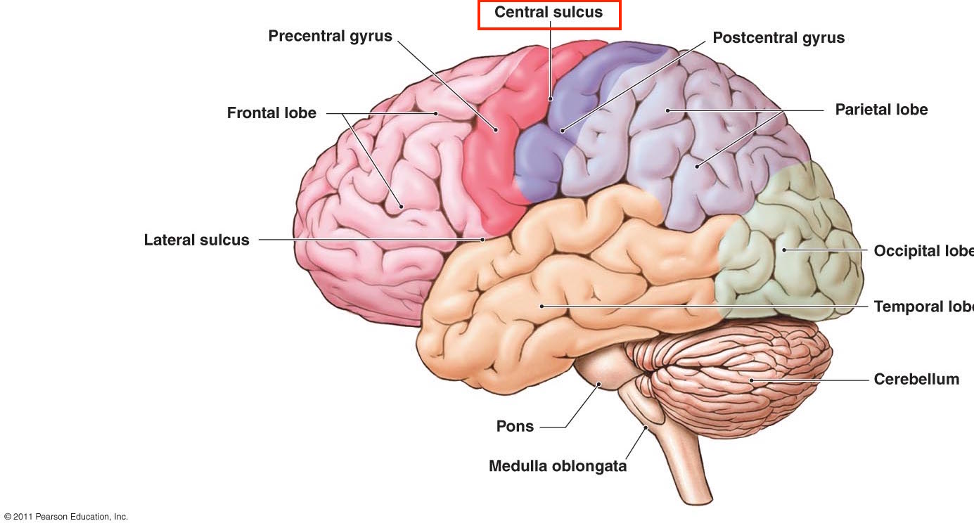
This page is dedicated to covering how the central sulcus will appear across different radiological studies. It is an important anatomical structure in the brain.
FINDING THE CENTRAL SULCUS ON AXIAL CROSS SECTIONS: OVERVIEW OF SIGNS
Given the importance of the central sulcus in orienting oneself to cranial anatomy, it is worth reviewing how one can quickly identify it. The following radiological signs are useful for finding the central sulcus on axial cross sections. The diagram below summarizes and depicts these different signs.
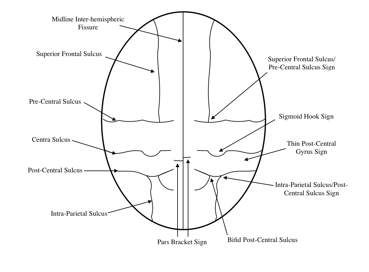
The sections below further discuss the signs that are useful to identifying the central sulcus on axial imaging of the brain.
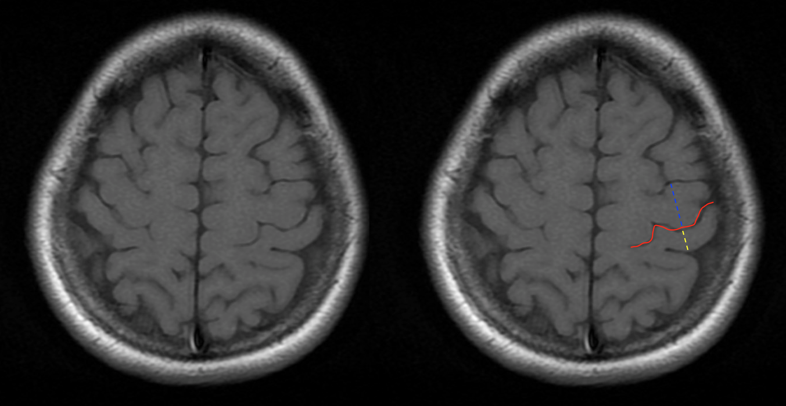
FINDING THE CENTRAL SULCUS ON AXIAL CROSS SECTIONS: SUPERIOR FRONTAL SULCUS SIGN (PRE-CENTRAL SULCUS SIGN)
Often times the posterior end of the frontal sulcus will connect to the pre-central sulcus. With this in mind, the central sulcus will reside more posterior to this sign.
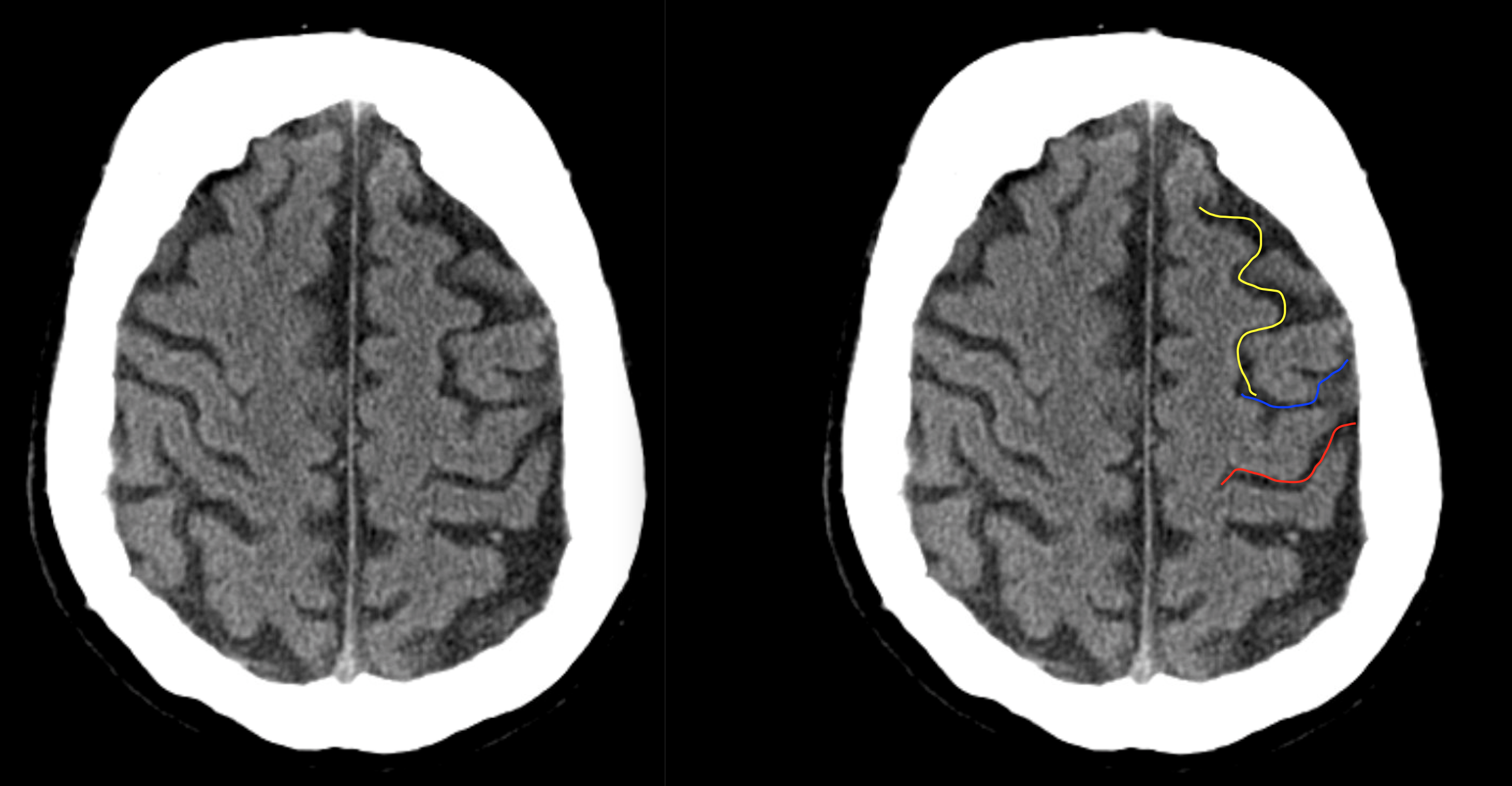
HERE ARE SOME EXAMPLES OF THIS SIGN SEEN ON A NON-CONTRAST HEAD CT-SCAN. CLICK THE THUMBNAILS TO VIEW THEM.


HERE ARE SOME EXAMPLES OF THIS SIGN SEEN ON A T1 WEIGHTED HEAD MRI WITHOUT CONTRAST. CLICK THE THUMBNAILS TO VIEW THEM.


FINDING THE CENTRAL SULCUS ON AXIAL CROSS SECTIONS: SIGMOID HOOK SIGN
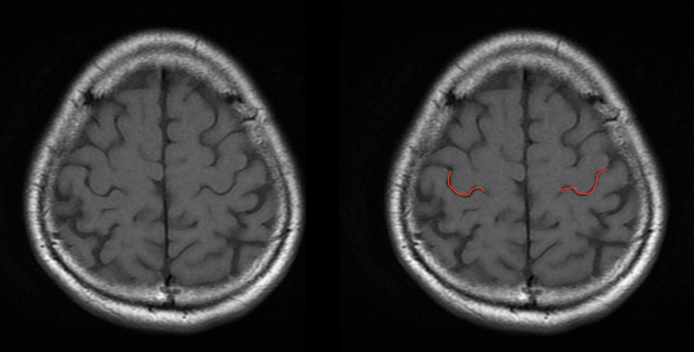
This “hook” refers to the shape of the sulcus that follows the posterior surface of the pre-central gyrus. This hook can be seen well most of the time on CT scans and is seen almost always on MRI. The area of the “hook” corresponds to the motor area of the pre-central gyrus that controls the hand.
HERE ARE SOME EXAMPLES OF THIS SIGN SEEN ON A NON-CONTRAST HEAD CT-SCAN. CLICK THE THUMBNAILS TO VIEW THEM.


HERE ARE SOME EXAMPLES OF THIS SIGN SEEN ON A T1 WEIGHTED HEAD MRI WITHOUT CONTRAST. CLICK THE THUMBNAILS TO VIEW THEM.


FINDING THE CENTRAL SULCUS ON AXIAL CROSS SECTIONS: PARS BRACKET SIGN
There are paired pars marginalia that form a “bracket” on either each side of the inter hemispheric fissure. These are at or posterior to the location of the central sulcus.
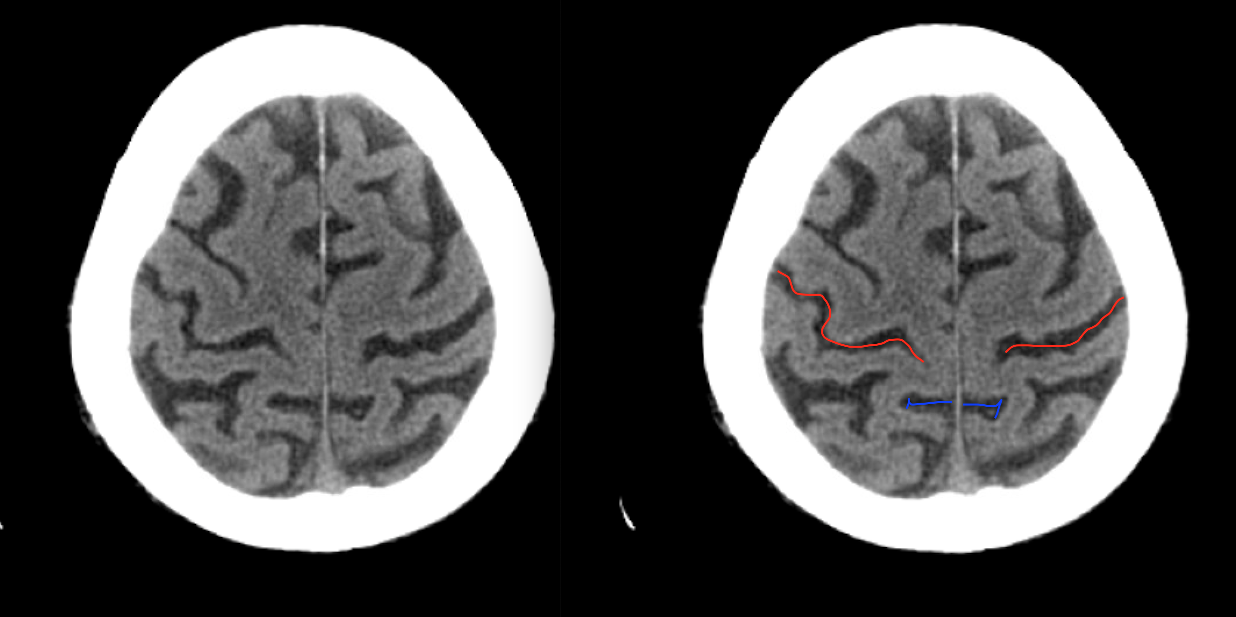
HERE ARE SOME EXAMPLES OF THIS SIGN SEEN ON A NON-CONTRAST HEAD CT-SCAN. CLICK THE THUMBNAILS TO VIEW THEM.


HERE ARE SOME EXAMPLES OF THIS SIGN SEEN ON A T1 WEIGHTED HEAD MRI WITHOUT CONTRAST. CLICK THE THUMBNAILS TO VIEW THEM.


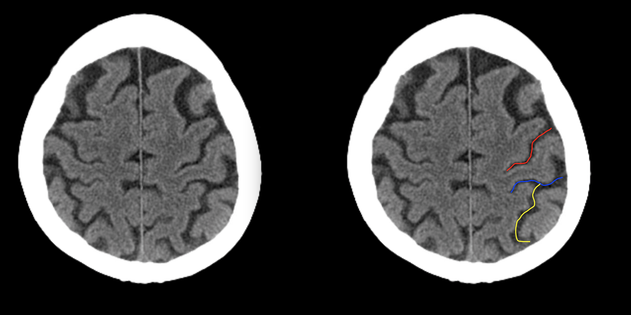
FINDING THE CENTRAL SULCUS ON AXIAL CROSS SECTIONS: BIFID POST-CENTRAL SULCUS SIGN
The post-central sulcus is often bifid in nature, and is located posterior to the central sulcus.

HERE ARE SOME EXAMPLES OF THIS SIGN SEEN ON A NON-CONTRAST HEAD CT-SCAN. CLICK THE THUMBNAILS TO VIEW THEM.


HERE ARE SOME EXAMPLES OF THIS SIGN SEEN ON A T1 WEIGHTED HEAD MRI WITHOUT CONTRAST. CLICK THE THUMBNAILS TO VIEW THEM.

FINDING THE CENTRAL SULCUS ON AXIAL CROSS SECTIONS: THIN POST-CENTRAL GYRUS SIGN
The post-central gyrus is typically thinner then the pre-central gyrus. This can help localize the central sulcus which sits in between these gyri.
HERE ARE SOME EXAMPLES OF THIS SIGN SEEN ON A NON-CONTRAST HEAD CT-SCAN. CLICK THE THUMBNAILS TO VIEW THEM.


HERE ARE SOME EXAMPLES OF THIS SIGN SEEN ON A T1 WEIGHTED HEAD MRI WITHOUT CONTRAST. CLICK THE THUMBNAILS TO VIEW THEM.


FINDING THE CENTRAL SULCUS ON AXIAL CROSS SECTIONS: INTRAPARIETAL SULCUS SIGN
The intraparietal sulcus will intersect the post central sulcus which can also be used to confirm the location of the central sulcus.
HERE ARE SOME EXAMPLES OF THIS SIGN SEEN ON A NON-CONTRAST HEAD CT-SCAN. CLICK THE THUMBNAILS TO VIEW THEM.


HERE ARE SOME EXAMPLES OF THIS SIGN SEEN ON A T1 WEIGHTED HEAD MRI WITHOUT CONTRAST. CLICK THE THUMBNAILS TO VIEW THEM.


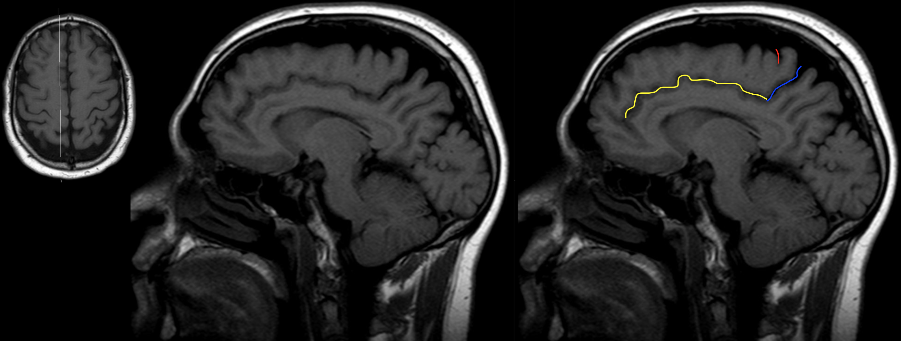
FINDING THE CENTRAL SULCUS ON SAGITTAL CROSS SECTIONS: CINGULATE SULCUS/PARS MARGINALIS SIGN
The central sulcus can also be appreciated on sagittal cross sections. Using the location of the cingulate suclcus and the pars marginalis can help localize the central sulcus. It is important to appreciate that the appearance and location of the central sulcus will vary depending upon where the sagittal section is anatomically. At sagittal sections close to the inter-hemispheric fissure, the central sulcus will be found immediately anterior to the pars marginalis. The pars marginalis can be identified relatively easily because it will be connected to the cingulate sulcus.
HERE ARE SOME EXAMPLES OF THIS SIGN SEEN ON A NON-CONTRAST HEAD CT-SCAN. CLICK THE THUMBNAILS TO VIEW THEM.


HERE ARE SOME EXAMPLES OF THIS SIGN SEEN ON A T1 WEIGHTED HEAD MRI WITHOUT CONTRAST. CLICK THE THUMBNAILS TO VIEW THEM.


ACKNOWLEDGEMENTS
A very special thanks goes to Dr. Pierre Sasson who made this page possible with his expertise and insight.
Page Updated: 11.15.2017