OVERVIEW
One of the other initial interpretations that needs to be made on a electrocardiogram (EKG/ECG) is in regards to the rhythm of the heart. The central question that is answered here is whether or not the heart beat originates from the sinus atrial node (i.e. sinus rhythm) or not.
Sinus rhythm refers to any cardiac rhythm where depolarization of the cardiac muscle begins at the sinus node, in the setting of alternating atrial and ventricular contraction. It is characterized by the presence of correctly-oriented P waves on the electrocardiogram (ECG). Sinus rhythm is necessary, but not sufficient, for normal electrical activity within the heart. In addition to correct P waves, the below flowchart can be used to help characterize what sinus rhythm is.
Reviewing the evaluating P waves page is helpful to understanding the criteria for sinus rhythm explained below.
CRITERIA FOR SINUS RHYTHM
Sinus rhythm is a term that can get unnecessarily complicated. All it means is that the origin of the heart beat is beginning at the sinus node (which is of course normal). In order to determine if a patient has sinus rhythm, one must ask themselves a few questions.
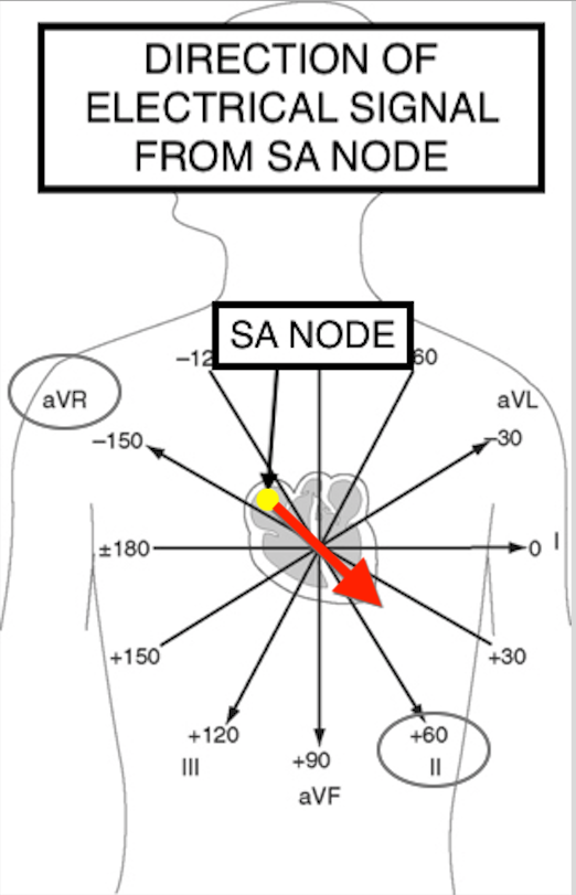
- Are there P waves present? Looking at leads I, II, and III p waves should be visible if the heartbeat is beginning at the SA node. If there are no P waves, then the patient does not have sinus rhythm.
- Is the P-wave/tracing positive in lead II? Anatomically, if the heartbeat begins at the SA node, it must be depolarizing toward lead II (making the tracing positive in this lead). If the tracing is negative in lead II the patient is not in sinus rhythm.
- Is the P-wave/tracing negative in lead AVR? Given that this lead is directly opposite lead II, if the patient has sinus rhythm, this tracing should be negative.
- Is there a p wave for every QRS complex and vice versa? Sinus rhythm requires alternating atrial and ventricular contractions.
EXAMPLES OF SINUS RHYTHM
Bellow is a gallery of examples of various leads with present P waves that originate from the SA node. This is an opportunity to being to appreciate how variable to quickly identify that a P wave is positive in lead II and negative in aVR for those who have P waves coming from the SA node. CLICK ON THE THUMBNAILS BELOW TO OPEN UP THIS IMAGE GALLERY.


Page Updated: 03.05.2018