OVERVIEW
This page covers exactly where electrocardiogram (EKG/ECG) leads are placed on the body. Understanding this placement can help the interpreting of EKG tracings down the line. It may be worth reading the page discussion how EKGs work as preparation for reading this page.
CHEST/PRE-CORDIAL LEADS
One must think of these leads as “cameras” that take electrical snapshots of how the heart is function from different perspectives around the body. If the electrical signal is going towards the lead a positive line is seen on the EKG, and if the signal is going away from the lead this is seen as negative on the EKG. There are two sets of 6 leads (chest and limb leads) that are used to generate axis around the heart.
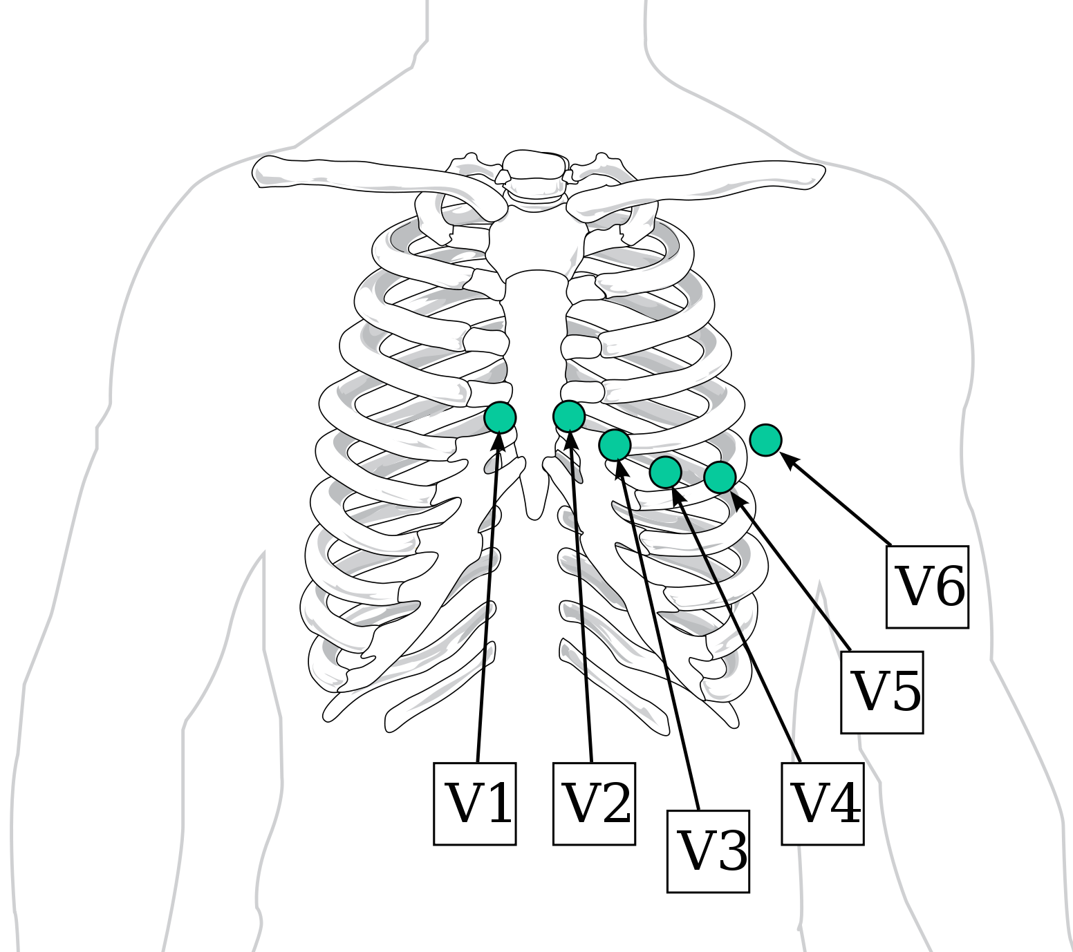
Chest/pre-coridal leads: these leads are arranged around the heart in an axial plane (think of a hula hoop around the chest). The leads are referred to as V1-V6 (see image below)
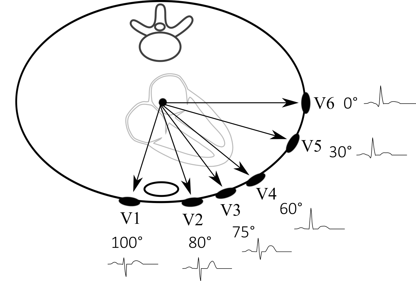
With the placement of these leads in mind, let us take a look at the position of the leads around the heart (below). Generally speaking leads next to the right side of the heart (i.e. V1) and leads next to the left side of the heart (i.e. V6) can be used to localize specific pathology (i.e. an enlarged left ventricle can increase the positivity of lead findings on leads such as V6 because more cardiac muscle is moving electrical activity towards that lead).
LIMB LEADS
The most important thing to keep in mind about these limb leads are that they form an axis around the heart that is perpendicular to the chest leads (if the chest leads are a hula hoop horizontally around the heart, the limb leads would be analogous to a vertical hula hoop that runs from the top to the bottom of the heart). Worrying about the specifics of each lead (i.e. which are unipolar vs. bipolar) is not terribly critical at this point, however keeping in mind their names and position (image below) is critical.
Generally speaking now we can appreciate the connection between leads and the inferior portion of the heart (II, III, AVF) , the right side of the heart (AVR), and the left side of the heart (AVL).
Page Updated: 03.04.2018