OVERVIEW
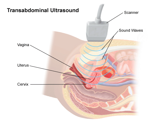
This page covers the important aspects of how to protocol (i.e. conduct) a transabdominal pelvic ultrasound. This is a commonly conducted study that has a clear protocol in place that will ensure all the proper images are taken. The various components of the study are outlined more below in detail, with example images. This exam is commonly performed with the transvaginal pelvic ultrasound protocol.
SUMMARY OF IMAGES/CLIPS THAT ARE NEEDED
Uterus:
- Sagittal View: image with entire body of uterus and cervix, same image with measurements of the longest sagittal axis and the corresponding perpendicular (AP) axis. Clip of entire uterus in this orientation.
- Transverse View: image at showing the transverse cross section of the uterus, same image measuring the transverse axis. Clip of entire uterus in this orientation.
Endometrium:
- Sagittal View: still image with the endometrium measured in the AP direction (perpendicular to long axis of endometrium).
Right Ovary/Right Adnexa:
- Sagittal View: image showing the ovary in this orientation. Clip of entire ovary in this orientation.
- Transverse View: image showing the ovary in this orientation. Clip of entire ovary in this orientation.
Left Ovary/Left Adnexa:
- Sagittal View: image showing the ovary in this orientation. Clip of entire ovary in this orientation.
- Transverse View: image showing the ovary in this orientation. Clip of entire ovary in this orientation.
Page Updated: 02.05.2018