Page Contents
OVERVIEW
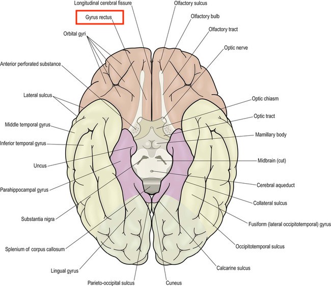
This page is dedicated to covering how the gyrus rectus will appear across different radiological studies. The gyrus rectus is a portion of the brain located at the medial most margin of the inferior surface of the frontal lobe. While its function is unclear, it may be involved in higher cognitive function (such as personality).
CLINICAL SIGNIFICANCE
The gyrus rectus is clinically significant because this portion of the brain is vulnerable to traumatic injury (hemorrhagic contusion). When going through scans of the brain it is important to look for olfactory groove meningiomas here and a focal non traumatic gyrus rectus hemorrhage should incite a search for an aneurysm of the anterior communicating artery.
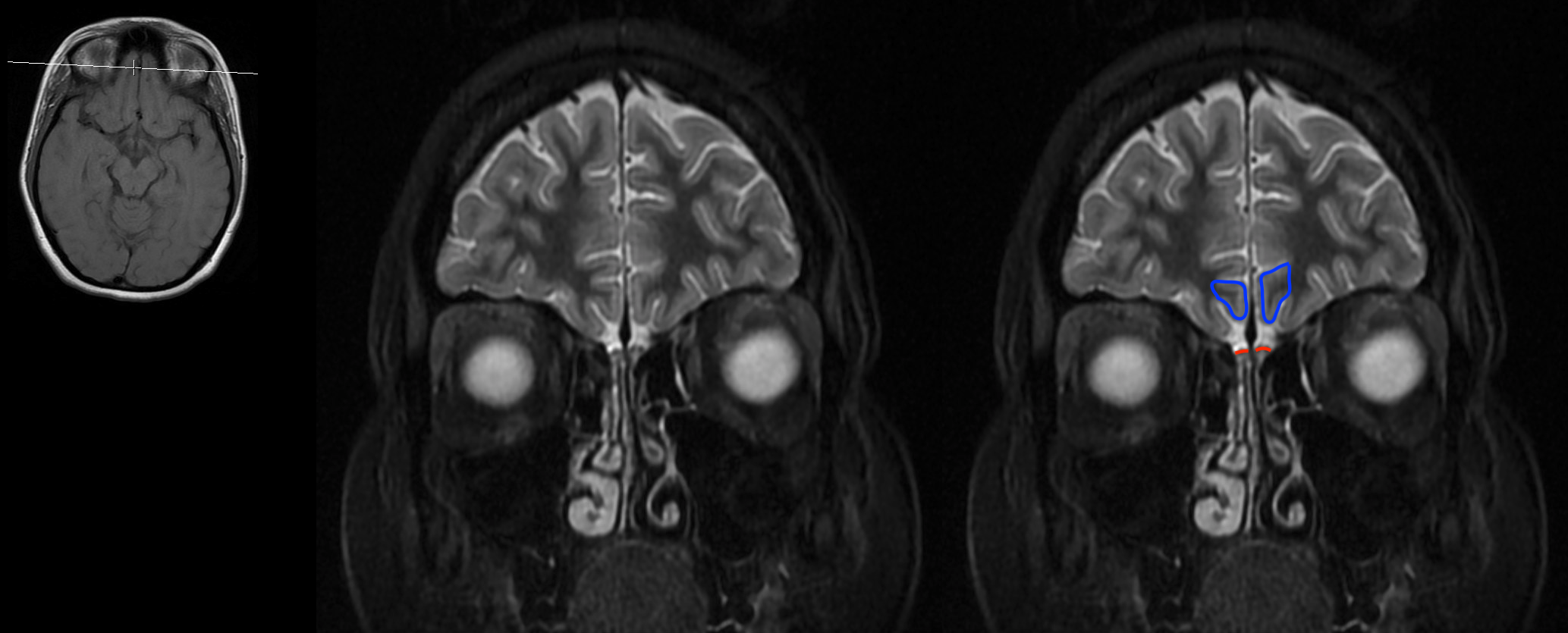
APPEARANCE OF THIS STRUCTURE ON CORONAL CROSS SECTIONS
The gyrus rectus can be seen on cross sectional imaging. To find the gyrus rectus on coronal cross sections it is useful to first find the olfactory nerve as it travels through the CSF filled olfactory groove. The gyrus rectus will be located directly above this nerve and will be the most medial gyrus.


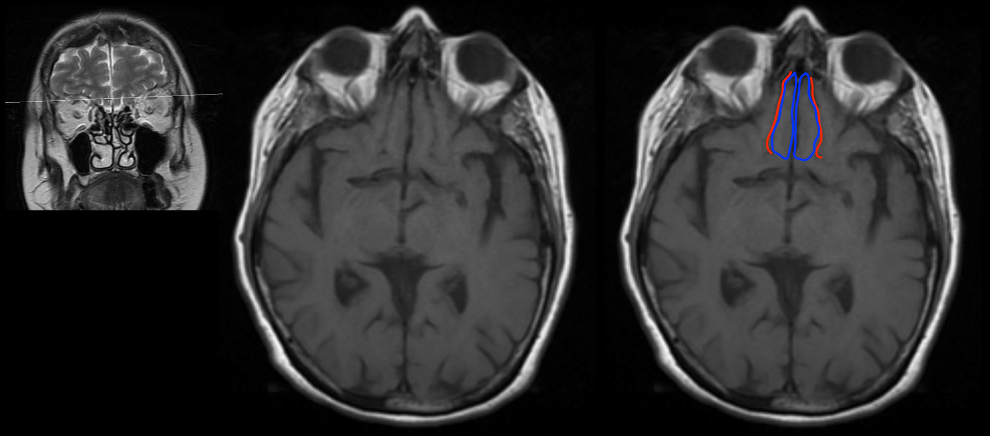
APPEARANCE OF THIS STRUCTURE ON AXIAL CROSS SECTIONS
The gyrus rectus can be seen on cross sectional imaging. The gyrus rectus on axial cross sections will be found immediately medial to the olfactory sulcus.
CLICK HERE FOR SOME EXAMPLES OF THIS STRUCTURE SEEN ON A T1 WEIGHTED HEAD MRI WITHOUT CONTRAST (AXIAL ORIENTATION)


ACKNOWLEDGEMENTS
A very special thanks goes to Dr. Pierre Sasson who made this page possible with his expertise and insight.
Page Updated: 01.22.2018