OVERVIEW
This page is dedicated to covering how the pars triangularis will appear across different radiological studies. It is an important anatomical structure in the brain.
APPEARANCE OF THE INFERIOR FRONTAL GYRUS ON SAGITTAL CROSS SECTIONS
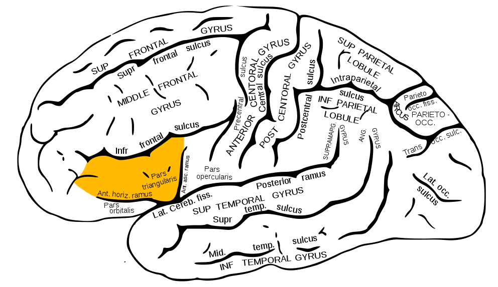
The pars triangularis can most easily be found by first identifying the inferior frontal gyrus (click on the link to learn how to identify this gyrus). As discussed on the inferior frontal gyrus page, this gyrus will have a characteristic “M” shape (referred to as the “M” sign. The pars orbitalis comprises the central portion of this gyrus (in other words, the middle “V” of the overall “M” shape). As another point of reference, the anterior horizontal ramus and the anterior ascending ramus will serve as borders for the pars triangularis and can be used to help identify its location. These rami come together to form the inferior tip of the pars triangularis and help to define its triangular shape (which is the origin of its name). The image below helps demonstrate these anatomical concepts.
HERE ARE SOME EXAMPLES OF THIS STRUCTURE SEEN ON A NON-CONTRAST HEAD CT-SCAN (SAGITTAL ORIENTATION). CLICK THE THUMBNAILS TO VIEW THEM.


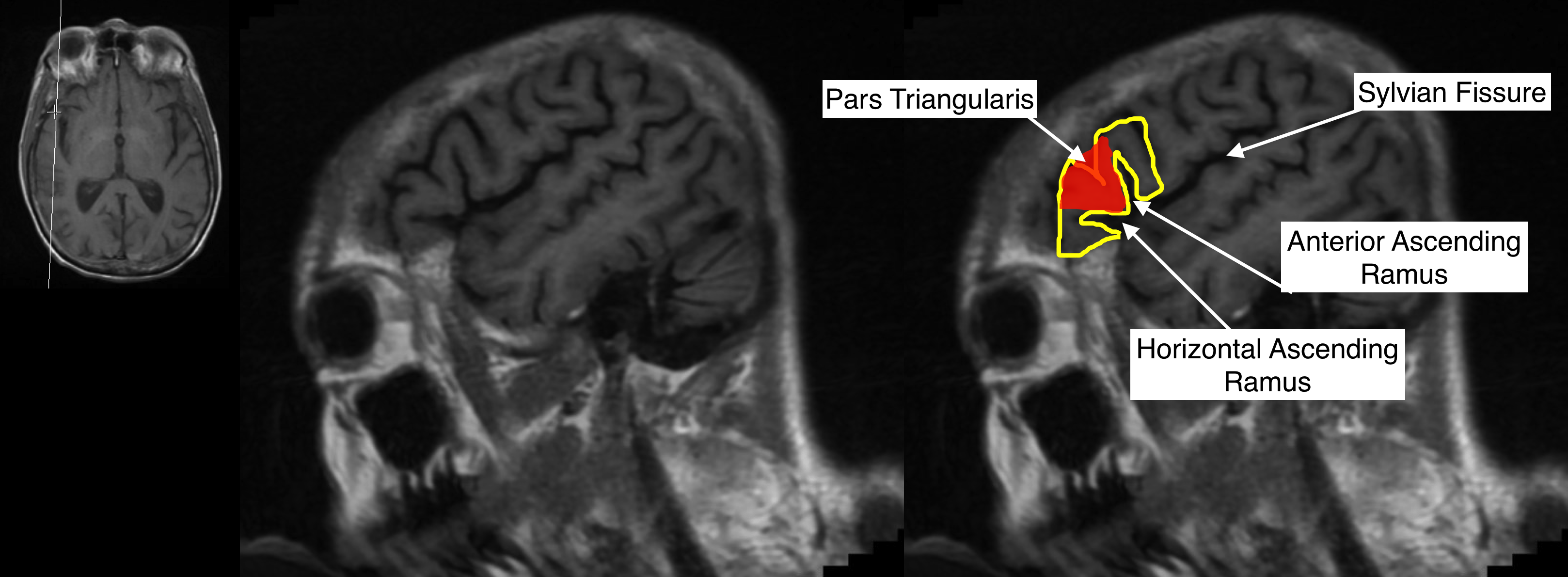
HERE ARE SOME EXAMPLES OF THIS STRUCTURE SEEN ON A T1 WEIGHTED HEAD MRI WITHOUT CONTRAST (SAGITTAL ORIENTATION). CLICK THE THUMBNAILS TO VIEW THEM.


ACKNOWLEDGEMENTS
A very special thanks goes to Dr. Pierre Sasson who made this page possible with his expertise and insight.
Page Updated: 12.25.2017