OVERVIEW
This page is dedicated to covering how the anterior horizontal rams of the Sylvian fissure will appear across different radiological studies. It is an important anatomical structure in the brain.
APPEARANCE OF THE HORIZONTAL ANTERIOR RAMUS OF THE SYLVIAN FISSURE ON SAGITTAL CROSS SECTIONS
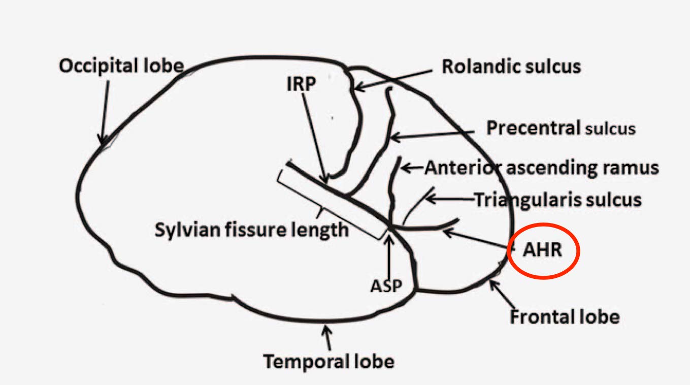
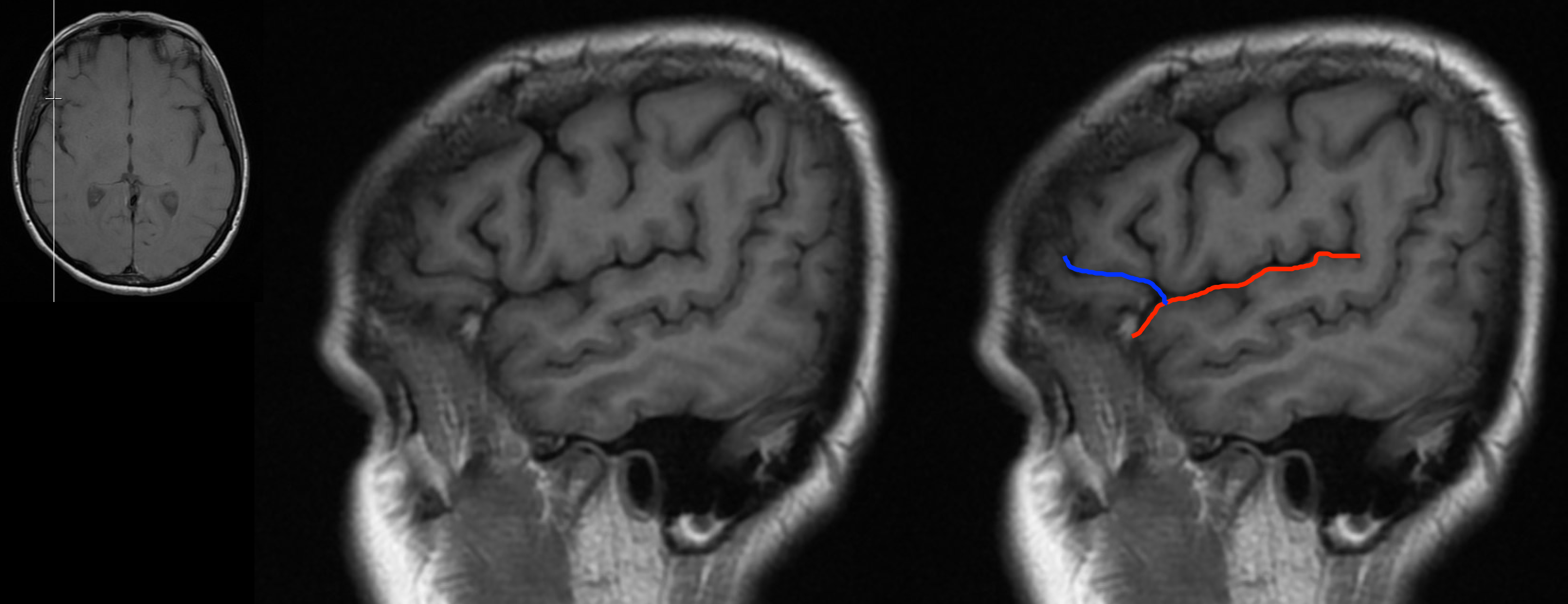
The horizontal anterior ramus of the Sylvian fissure can be fond on sagittal sections by first identifying the Sylvian fissure. This ramus will originate from the anterior aspect of the Sylvian fissure and extends horizontally (as demonstrated in the image below).
HERE ARE SOME EXAMPLES OF THIS RAMUS SEEN ON A NON-CONTRAST HEAD CT-SCAN (SAGITTAL ORIENTATION). CLICK THE THUMBNAILS TO VIEW THEM.


HERE ARE SOME EXAMPLES OF THIS RAMUS SEEN ON A T1 WEIGHTED HEAD MRI WITHOUT CONTRAST (SAGITTAL ORIENTATION). CLICK THE THUMBNAILS TO VIEW THEM.


ACKNOWLEDGEMENTS
A very special thanks goes to Dr. Pierre Sasson who made this page possible with his expertise and insight.
Page Updated: 12.24.2017