Page Contents
OVERVIEW
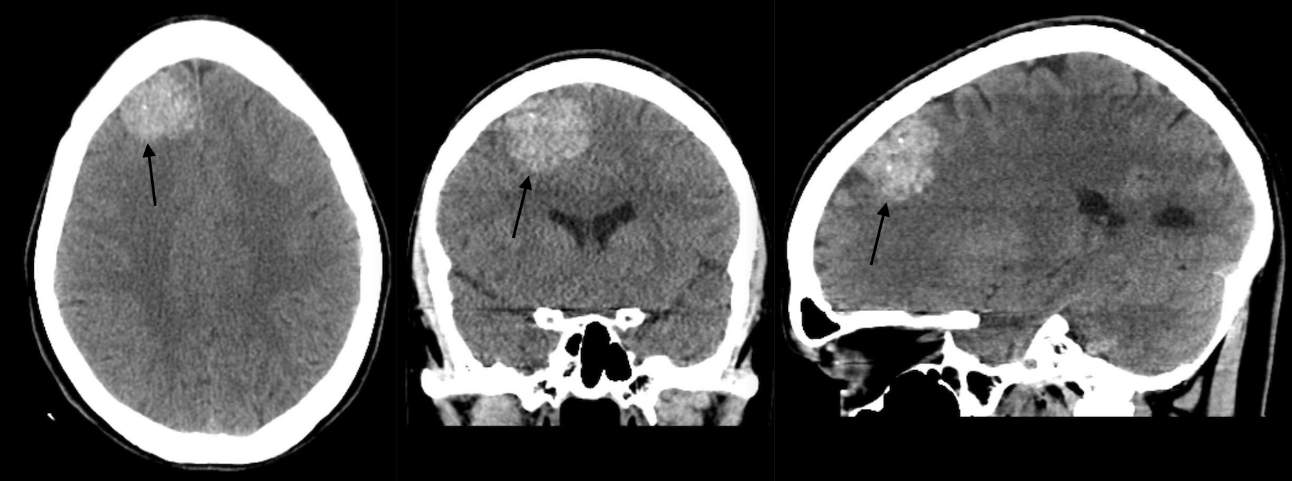
This page is dedicated to covering the important radiological finding of increased signal attenuation in the brain (0n a non-contrast head CT-scan).
WHAT IS IT?
Increased signal attenuation refers to a finding on CT studies where a tissue/structure appears to “more white” for a certain reason. In the case of a non-contrast CT-scan this reason will be due to an increased density of the structure in question.
DIFFERENTIAL DIAGNOSIS FOR THIS FINDING
When seeing a increased signal attenuation in the brain (0n a non-contrast head CT-scan), it is important to keep in mind the following possible causes of this finding:
- Bleeding
- Neoplasm
- Vascular malformations
KEY FEATURES TO LOOK FOR WHEN CHARACTERIZING THE FINDING
When seeing a increased signal attenuation in the brain (0n a non-contrast head CT-scan), there are a few important radiological features one should look at to try and characterize the finding. These features can help navigate the differential diagnosis above.
- Location: is the mass inside or outside the brain parenchyma (intraaxial vs. extraxial?)
Page Updated: 08.23.2017