Page Contents
OVERVIEW
This page is dedicated to covering how the condition dural arteriovenous fistula will appear on different types of imaging studies.
BASIC CHARACTERISTICS
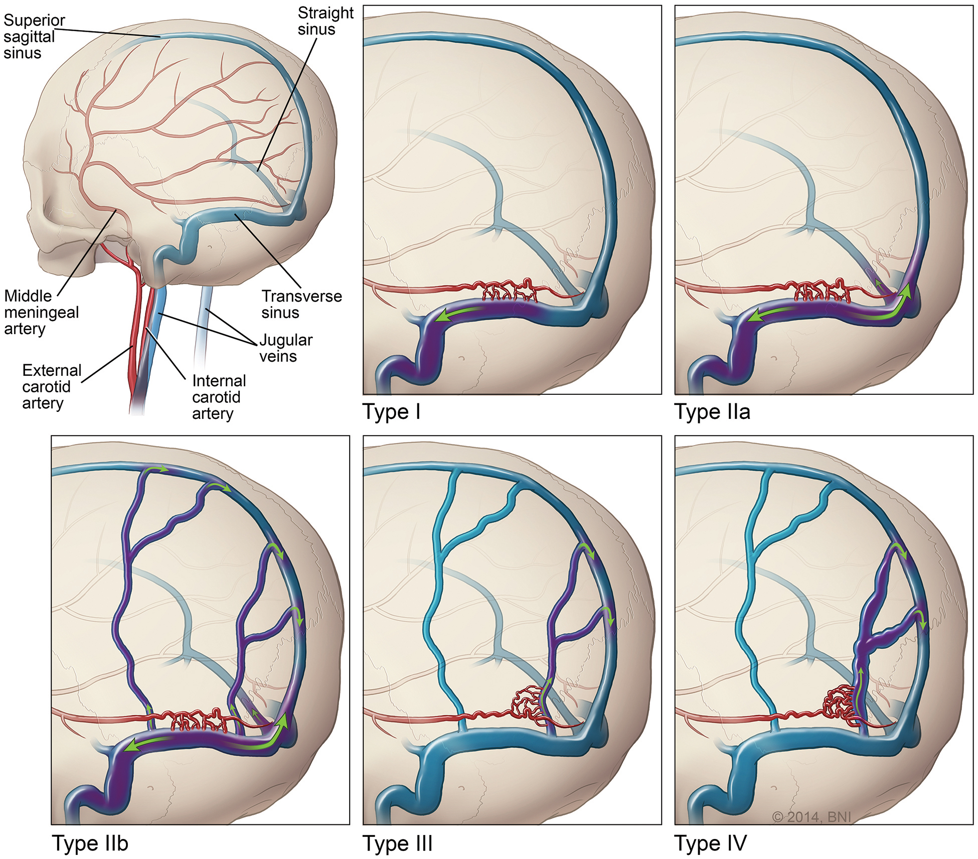
Fundamentally, a dural arteriovenous fistula refers to an abnormal connection between the branches of dural arteries to dural veins or a venous sinus.
Here are some general features of this condition that might be appreciated across modalities:
- Abnormally enlarged/tortuous vessels might be present in the subarachnoid space
- Arterialization of venous sinuses during arterial phase of contrast studies
COMPUTERIZED TOMOGRAPHY (CT-SCAN)
Key features of the appearance of this condition on this imaging modality are:
MAGNETIC RESONANCE IMAGING (MRI)
Key features of the appearance of this condition on this imaging modality are:
Page Updated: 08.09.2017