Page Contents
OVERVIEW
This page is dedicated to discussing the various different types of MRI artifacts that can occur.
- Gibbs (truncation) artifact
- Ghosting (Nyquist, or N/2) artifact
- Chemical shift artifact
- Zipper artifacts
- Spike noise artifacts
- Aliasing (“wrap-around”) artifact
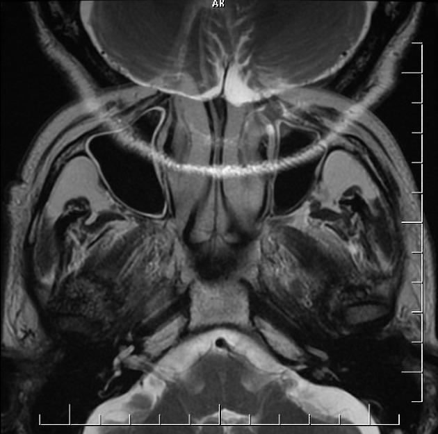
GIBBS ARTIFACT
What is it?
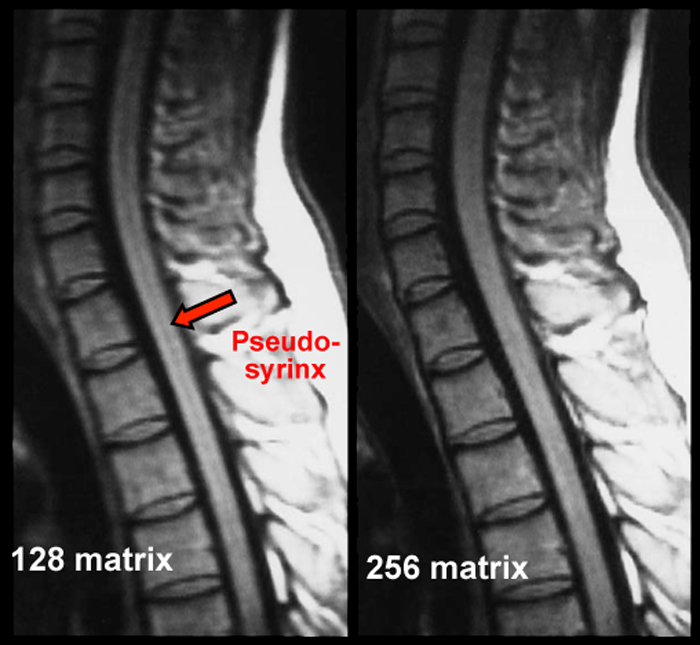
A Gibbs (truncation) artifact occurs as a consequence of using Fourier transforms to reconstruct MR signals into representative images. It appears as multiple fine parallel lines that are immediately adjacent to high contrast interfaces.
Not only can this artifact be distracting, it can also mimic the appearance of a syrinx in the spinal cord which can be problematic clinically.
How to solve it: This artifact can be resolved by
- Specifying more phase encodings
- Awaiting more samples during frequency encoding (increased matrix)
- Pre/post reconstruction image filtration.
GHOSTING (NYQUIST, OR N/2) ARTIFACT
What is it?
A ghosting (Nyquist, or N/2) artifact is most often caused by some type of motion and appears as an image copy/multiple copies of an image that can blur the image.
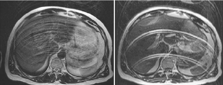
How to solve it: This artifact can be resolved by
- Having patients hold still
- Having patient’s hold breath
- Pharmaceuticals to reduce bowel motion.
CHEMICAL SHIFT ARTIFACT
What is it?
A chemical shift artifact will appear as a bright or black band at the interfaces of fatty tissue/non-fatty tissue. The bright band will be present on the side of the fatty tissue, and the black band will be present on the non-fatty tissue. This is caused by the resonant frequency of fat that mimics frequency-encoding.
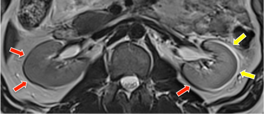
How to solve it: This artifact can be resolved by
- Inverting phase-encode and frequency-encode directions to view pathology in an improved fashion
ZIPPER ARTIFACTS
What is it?
These appear as a strip of noise in an image. These are caused by extraneous RF signals near the MRI machine during readout.
How to solve it: This artifact can be resolved by
- Making sure door to scanner room is closed
- Make sure equipment in room is checked for RF emissions (such as contrast pumps etc).
- Call medical physics staff/field engineer to find RF leak.
SPIKE NOISE ARTIFACT
What is it?
Series of bands alternating between light and dark bands superimposed over an image.
How to solve it: This artifact can be resolved by
- Calling field engineer to check for vibrating metal/loose wires in MRI machine
ALIASING (“WRAP-AROUND”) ARTIFACT
What is it?
An aliasing (“wrap-around) artifact is when an structure (outside the field of view) wraps around the image on the side opposite it it’s anatomical location.
How to solve it: This artifact can be resolved by
- Orient the thinner axis of anatomy along the phase-encoding direction
- Enlarge the field of view
- Exclude all anatomy from the top view slices (for 3D ahead exams)
- Use spatial saturation bands to null tissues outside the field of view.
ARTIFACT
What is it?
How to solve it: This artifact can be resolved by
Page Updated: 08.06.2017