Page Contents
OVERVIEW
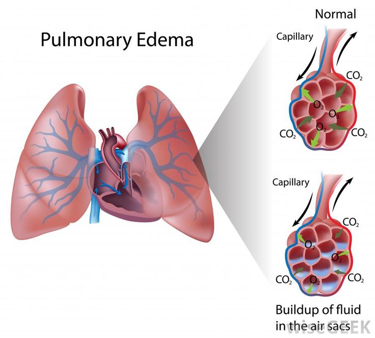
This page is dedicated to covering how the condition alveolar pulmonary edema will appear on different types of radiological imaging studies. Other pages dedicated to the radiological appearance of conditions can be found here.
BASIC CHARACTERISTICS
Fundamentally alveolar pulmonary edema refers to the edema/fluid collection in the alveoli of the lung. Typically this condition is caused by cardiac issues (most classically left heart failure). By definition, alveolar pulmonary edema is an airspace disease.
Here are some features of pleural effusion that can be observed across all imaging studies:
- Increased opacity/signal: the nature of this condition is that fluid will fill the alveoli. This can appear as an opacity/increased signal on different studies.
- Location: most of the time alveolar pulmonary edema will be bilateral in nature (however it may be asymmetric).
- Associated conditions: pleural effusions are often times associated with alveolar pulmonary edema that is cardiac in origin.
X-RAY
A chest X-ray can be a very common study by which alveolar pulmonary edema can be appreciated. Make sure to review how to read chest X-rays, as well as the archive of unremarkable chest X-rays as a reference point.
Some key features to keep in mind for the appearance of alveolar pulmonary edema on a chest X-ray are:
- Presence of opacity: fundamentally the fluid that collects in the airspace can result in the presence of hazy opacities.
- Bilateral nature: typically pulmonary edema is not a unilateral condition and affects both lungs.
- Configuration (perihilar): acute pulmonary edema typically affects the perihilar airspaces bilaterally (and causes a characteristic “bat wing” or “angel wing” configuration.
- Absence of air bronchograms: typically (unlike other airway diseases) pulmonary edema does not produce air bronchograms radiologically. This is because fluid fills not only the iarpsaces but the bronchi as well in this condition. They may still be present however it is less of a classic presentation and the presence of air bronchograms DOES NOT RULE OUT the diagnosis of pulmonary edema.
- Quick resolution following treatment: patients treated for pulmonary edema should see improvement < 48 hours on repeat X-rays.
COMPUTIRIZED TOMOGRAPHY (CT-SCAN)
MAGNETIC REASONANCE IMAGING (MRI)
Page Updated: 01.07.2016