Page Contents
OVERVIEW
This page is dedicated to covering how a generalized ileus will appear on different types of radiological imaging studies.
BASIC CHARACTERISTICS
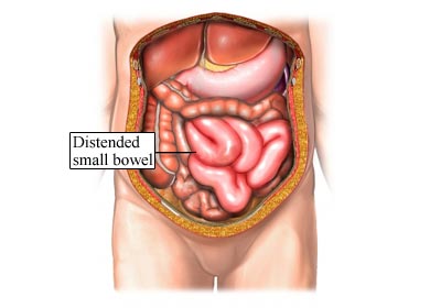
Fundamentally an ileus refers to the atony (loss of tone) of part/or all of the intestines. In the case of a generalized ileus many bowel loops will be involved in this atonic process. This means that the bowels will be unable to contract normally and move waste out of the body. While there can be a number of different causes of ileus, the shared features of what a ileus actually is helps with radiological analysis.
Here are some features of a ileus that can be observed across all imaging studies:
- Location: typical it is the small bowel that is involved in this process.
- Dilated bowel loops: the normal intestine is < 2.5 cm in diameter. This diameter will often be increased in portions of the small intestine involved in the localized ileus. It is important to note that these loops do not dilate as much as ones seen in an obstructive process (like a small bowel obstruction).
SUPINE ABDOMINAL X-RAY (KUB)
An abdominal X-ray can be one of the first studies ordered when a ileus is suspected. The supine view will be one of the images collected.
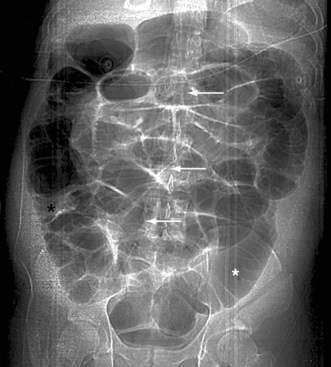
Some key features to keep in mind for the appearance of a localized ileus on a supine KUB
- Location: both the small and large intestines can be involved.
- Amount of dilated bowel loops: given that the condition is a generalized ileus, many bowel loops will be involved and they will be visibly dilated.
- Gas in sigmoid/rectum: often gas will be present in the sigmoid colon or rectum, because it can move past the localized ileus.
UPRIGHT ABDOMINAL X-RAY (KUB)
An abdominal X-ray can be one of the first studies ordered when a ileus is suspected. The supine view will be one of the images collected.
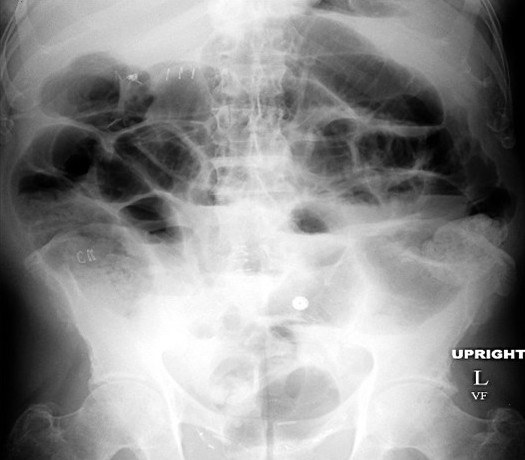
Some key features to keep in mind for the appearance of a localized ileus on a supine KUB
- Location: both the small and large intestines can be involved.
- Amount of dilated bowel loops: given that the condition is a generalized ileus, many bowel loops will be involved and they will be visibly dilated.
- Gas in sigmoid/rectum: often gas will be present in the sigmoid colon or rectum, because it can move past the localized ileus.
COMPUTIRIZED TOMOGRAPHY (CT-SCAN)
MAGNETIC REASONANCE IMAGING (MRI)
Page Updated: 10.08.2016