Page Contents
OVERVIEW
This page is dedicated to covering how the condition atelectasis will appear on different types of radiological imaging studies.
BASIC CHARACTERISTICS
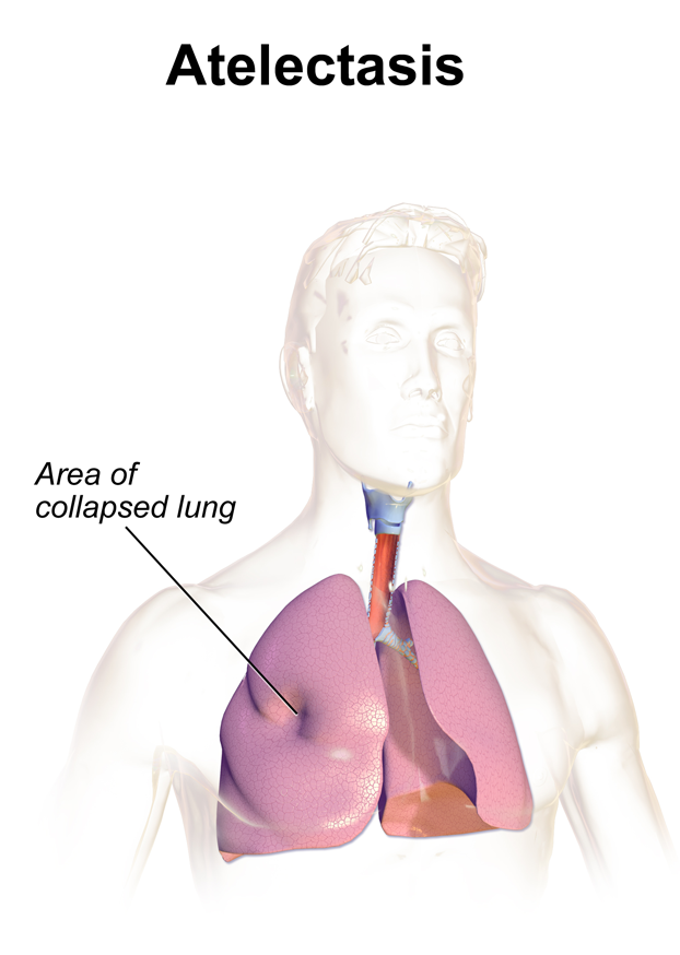
Fundamentally, atelectasis refers to the collapse of the lung parenchyma WITH the qualifier that the visceral and parietal pleura remain attached. This is different from other types of lung collapse such as pneumothorax (where air enters between the parietal and visceral pleura, separating them).
There are multiple causes of atelectasis that fall into the following categories:
- Obstructive: this type of collapse is caused by some process that obstructs airflow out of a region of the lung (mucus plug, tumor, etc).
- Compressive: a mass effect near by (such a s pleural effusion or a pneumothorax) will compress the lung parenchyma and cause it to collapse.
- Cicatrization: scarring in a nearby region will result in the collapse of a lung region.
Here are some general features of atelectasis:
- Opacity/increased signal: collapse of the lung parenchyma will increase the density in the affected area, resulting in an opacity on a chest X-ray (or increased signal on other studies).
- Shift of mediastinal structures TOWARD the atelectasis: given that the parietal and visceral pleura remain intact with one another, the vacuum from the collapse will pull nearby structures toward the atelectasis.
There are many different types of atelectasis: essentially any amount of the lung tissue can be involved in this process, which can manifest a few different ways.
- Complete atelectasis (causing hemithorax opacification): complete atelectasis of a lung can be responsible for causing a complete “white-out” over a lung field on a chest X-ray.
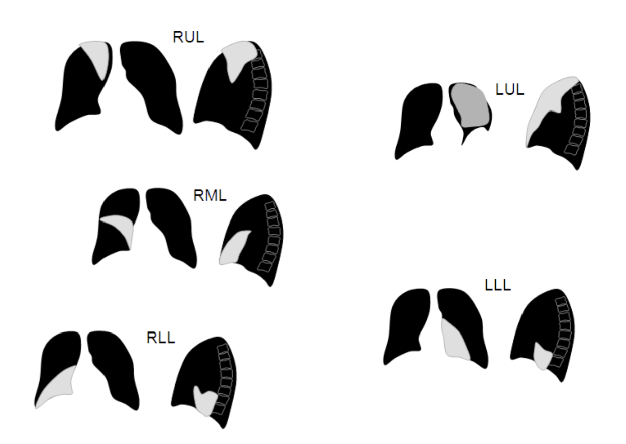
- Lobar atelectasis: given the nature of this condition, any of the 5 lung lobes can be affected.
CHEST X-RAY
A chest X-ray can be a very common study by which atelectasis will be diagnosed. Make sure to review how to read chest X-rays, as well as the archive of unremarkable chest X-rays as a reference point.
Some key features to keep in mind for the appearance of atelectasis on a chest X-ray are:
- Presence of opacity: fundamentally the collapse of this lung parenchyma will appear as an opacity over a portion of the lung field. On a chest X-ray this will show up as “increased white”.
- Sharp margins: generally the portions of collapsed lung tissue will have sharp margins that can be appreciated radiographically on an X-ray.
- Mediastinal shift toward the opacification: in certain cases the lung collapse will cause the shift of mediastinal structures toward the atelectasis (due to “vacuum” generated by the collapse of this lung tissue). This will be more obvious in larger collapses (such as complete atelectasis).
RIGHT UPPER LOBE ATELECTASIS
The archive below organizes different examples of a right upper lobe atelectasis. It will really be the anatomical location of the finding that will help diagnosis this SPECIFIC subtype of atelectasis. Click on the thumbnails below to view the archive.
 Example 1
 Example 2


RIGHT MIDDLE LOBE ATELECTASIS
The archive below organizes different examples of a left middle lobe atelectasis. It will really be the anatomical location of the finding that will help diagnosis this SPECIFIC subtype of atelectasis. Whats more, this type of atelectasis often obscures the right cardiac border. Click on the thumbnails below to view the archive.
 Example 1
 Example 2


RIGHT LOWER LOBE ATELECTASIS
The archive below organizes different examples of a right lower lobe atelectasis. It will really be the anatomical location of the finding that will help diagnosis this SPECIFIC subtype of atelectasis.Whats more, this type of atelectasis often obscures the border of the right diaphragm. Click on the thumbnails below to view the archive.
 Example 1
 Example 2


LEFT UPPER LOBE ATELECTASIS
The archive below organizes different examples of a left upper lobe atelectasis. It will really be the anatomical location of the finding that will help diagnosis this SPECIFIC subtype of atelectasis. Click on the thumbnails below to view the archive.
 Example 1
 Example 2


LEFT LOWER LOBE ATELECTASIS
The archive below organizes different examples of a left lower lobe atelectasis. It will really be the anatomical location of the finding that will help diagnosis this SPECIFIC subtype of atelectasis. Click on the thumbnails below to view the archive.
 Example 1
 Example 2


COMPLETE ATELECTASIS
This archive below organizes different examples of complete atelectasis. This will result in a complete white out of the affected lung. Make sure to review the page discussing this radiological finding of complete lung “white out” called hemithorax opacification. Click on the thumbnails below to view the archive.
 Example 1
 Example 2


COMPUTIRIZED TOMOGRAPHY (CT-SCAN)
Atelectasis can also be visualized on a CT scan
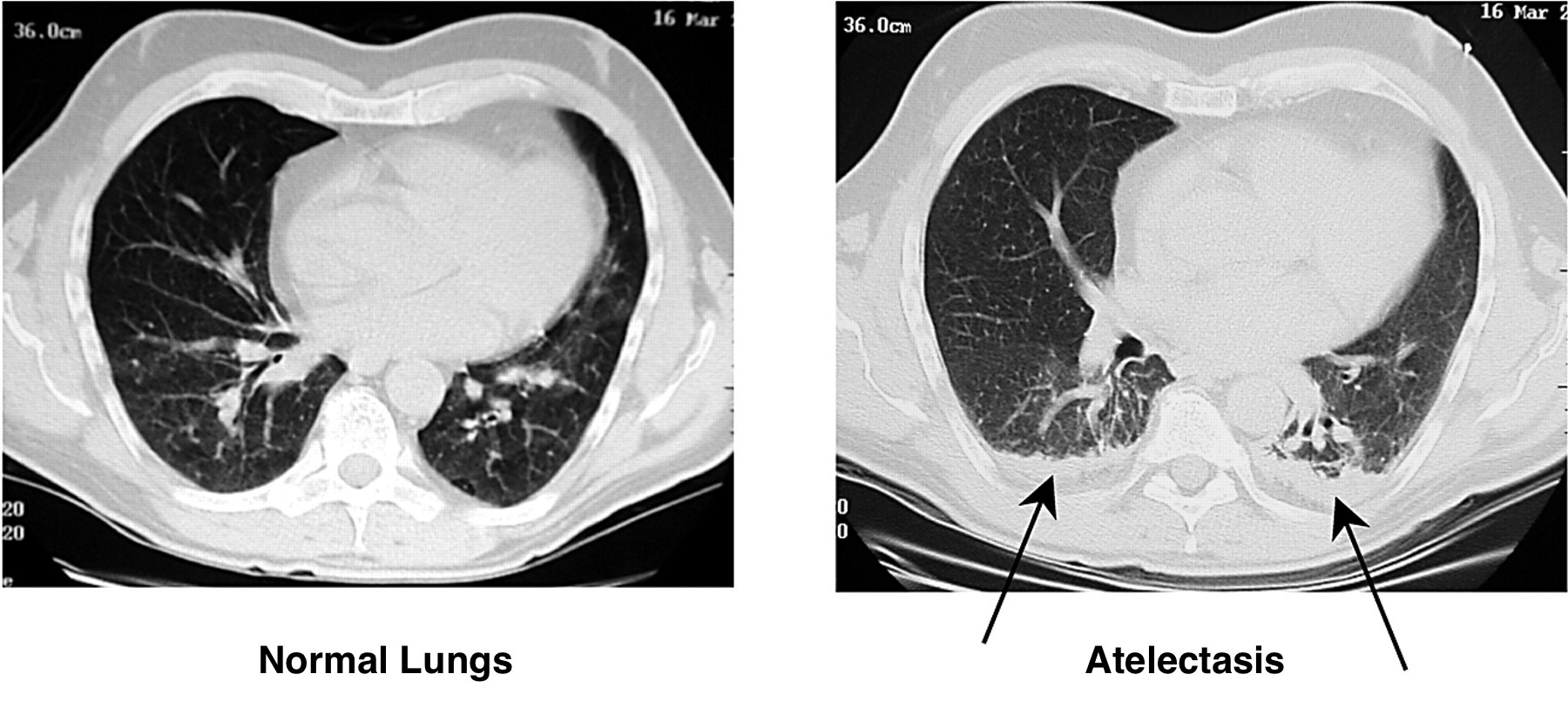
Some key features to keep in mind for the appearance of atelectasis on a CT-scan are:
- Presence of signal: fundamentally the collapse of this lung parenchyma will appear over a portion of the lung field. On a CT- scan this will show up as an increased signal.
MAGNETIC REASONANCE IMAGING (MRI)
Atelectasis can also be visualized on an MRI
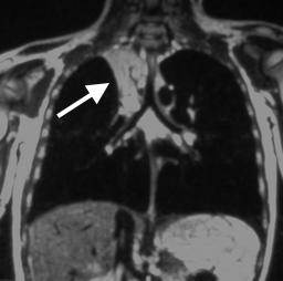
Some key features to keep in mind for the appearance of atelectasis on an MRI are:
- Presence of signal: fundamentally the collapse of this lung parenchyma will appear over a portion of the lung field. On an MRI this will show up as an increased signal (specific type of signal will depend on the type of study).
Page Updated: 10.11.2016